. 1998 Sep;36(9):2671-80.
doi: 10.1128/JCM.36.9.2671-2680.1998.

Cloning and characterization of multigenes encoding the immunodominant 30-kilodalton major outer membrane proteins of Ehrlichia canis and application of the recombinant protein for serodiagnosis

Affiliations

Cloning and characterization of multigenes encoding the immunodominant 30-kilodalton major outer membrane proteins of Ehrlichia canis and application of the recombinant protein for serodiagnosis

N Ohashi et al. J Clin Microbiol. 1998 Sep.

Abstract

A 30-kDa major outer membrane protein of Ehrlichia canis, the agent of canine ehrlichiosis, is the major antigen recognized by both naturally and experimentally infected dog sera. The protein cross-reacts with a serum against a recombinant 28-kDa protein (rP28), one of the outer membrane proteins of a gene (omp-1) family of Ehrlichia chaffeensis. Two DNA fragments of E. canis were amplified by PCR with two primer pairs based on the sequences of E. chaffeensis omp-1 genes, cloned, and sequenced. Each fragment contained a partial 30-kDa protein gene of E. canis. Genomic Southern blot analysis with the partial gene probes revealed the presence of multiple copies of these genes in the E. canis genome. Three copies of the entire gene (p30, p30-1, and p30a) were cloned and sequenced from the E. canis genomic DNA. The open reading frames of the two copies (p30 and p30-1) were tandemly arranged with an intergenic space. The three copies were similar but not identical and contained a semivariable region and three hypervariable regions in the protein molecules. The following genes homologous to three E. canis 30-kDa protein genes and the E. chaffeensis omp-1 family were identified in the closely related rickettsiae: wsp from Wolbachia sp. , p44 from the agent of human granulocytic ehrlichiosis, msp-2 and msp-4 from Anaplasma marginale, and map-1 from Cowdria ruminantium. Phylogenetic analysis among the three E. canis 30-kDa proteins and the major surface proteins of the rickettsiae revealed that these proteins are divided into four clusters and the two E. canis 30-kDa proteins are closely related but that the third 30-kDa protein is not. The p30 gene was expressed as a fusion protein, and the antibody to the recombinant protein (rP30) was raised in a mouse. The antibody reacted with rP30 and a 30-kDa protein of purified E. canis. Twenty-nine indirect fluorescent antibody (IFA)-positive dog plasma specimens strongly recognized the rP30 of E. canis. To evaluate whether the rP30 is a suitable antigen for serodiagnosis of canine ehrlichiosis, the immunoreactions between rP30 and the whole purified E. canis antigen were compared in the dot immunoblot assay. Dot reactions of both antigens with IFA-positive dog plasma specimens were clearly distinguishable by the naked eye from those with IFA-negative plasma specimens. By densitometry with a total of 42 IFA-positive and -negative plasma specimens, both antigens produced results similar in sensitivity and specificity. These findings suggest that the rP30 antigen provides a simple, consistent, and rapid serodiagnosis for canine ehrlichiosis. Cloning of multigenes encoding the 30-kDa major outer membrane proteins of E. canis will greatly facilitate understanding pathogenesis and immunologic study of canine ehrlichosis and provide a useful tool for phylogenetic analysis.


Figures

FIG. 1
FIG. 1
Genomic Southern blot analysis of E. canis DNA with the partial p30 gene probe (A) and with the partial p30a gene probe (B) and schematic representation of the blotting patterns (C). Numbers indicate molecular sizes in kilobases. Filled dots, bands hybridized with both p30 and p30a probes; striped dots, bands hybridized with p30a probe alone; lightly shaded dots, bands hybridized with p30 probe alone.
FIG. 2
FIG. 2
Amino acid sequence alignment of P30, P30-1, and P30a of E. canis, seven members of E. chaffeensis omp-1 multigene family (P28 and OMP-1A to OMP-1F), and MAP-1 of C. ruminantium (Senegal strain). The sequences of the E. chaffeensis omp-1 gene family and MAP-1 are from the reports of Ohashi et al. (22) and Van Vliet et al. (31), respectively. Aligned positions of identical amino acids with P30 of E. canis are indicated by dots. Gaps (indicated by dashes) were introduced for optimal alignment of all proteins. Bars indicate an SV and three HVs (HV1, -2, and -3). The arrowhead indicate the putative cleavage site of the signal peptide.
FIG. 3
FIG. 3
Phylogenetic classification among P30, P30-1, and P30a of E. canis and the major OMPs of the closely related rickettsiae based on amino acid sequence similarities. Evolutionary distance values were determined by the method described by Kimura, and the tree was constructed by the unweighted pair-group method of analysis. Scale bar indicates 10% divergence in amino acid sequences. Bootstrap values from 100 analyses are shown at the branch points of the tree. Bars with symbols indicate representative clusters. The GenBank accession numbers of the major OMP gene sequences of the organisms used in the analysis are as follows: P28 (E. chaffeensis), U72291; OMP-1B to OMP-1F (E. chaffeensis), AF021338; MAP-1 (C. ruminantium Senegal strain), I40882, MAP-1 (C. ruminantium Antigua strain), U50830; MAP-1 (C. ruminantium Gardel strain), U50832; MAP-1 (C. ruminantium Um Banein strain), U50835; MAP-1 (C. ruminantium Nyatsanga strain), U50834; MAP-1 (C. ruminantium Welgevonden strain), U49843; MAP-1 (C. ruminantium Crystal Springs strain), U50831; MAP-1 (C. ruminantium Highway strain), U50833; WSP (Wolbachia sp. Wha strain), AF020068; WSP (Wolbachia sp. Wcof strain), AF020067; WSP (Wolbachia sp. WmelH strain), AF020066; WSP (Wolbachia sp. Wri strain), AF020070; MSP-4 (A. marginale), Q07408; MSP2-1 (A. marginale), U07862; MSP2-2 (A. marginale), U36193; and P44 (HGE agent), AF059181.
FIG. 4
FIG. 4
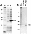
SDS-PAGE profiles of a recombinant clone expressing P30 of E. canis (A) and the purified recombinant protein (B). Gels were stained with Coomassie blue. Lanes: M, molecular size markers; C, pET29-transformed E. coli (negative control); R, pET29p30-transformed E. coli (recombinant); Eca, purified E. canis; PP-rP30, partially purified rP30 fusion protein of E. canis; and AP-rP30, affinity-purified rP30 fusion protein. The recombinant rP30 protein is indicated by the arrow. The numbers on the left of each panel indicate molecular masses in kilodaltons.
FIG. 5
FIG. 5
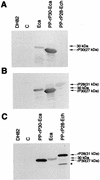
Western blot analysis with clinical dog plasma with canine ehrlichiosis (A and B) and mouse anti-rP30 serum (C). (A) Dog plasma with a 1:40 IFA titer against E. canis; (B) dog plasma with a 1:1,280 IFA titer. Lanes: DH, DH82 dog macrophage cell (negative control); C, a pET29-transformed E. coli (negative control); Eca, purified E. canis (reactive 30-kDa protein is indicated by arrows in each panel); PP-rP30-Eca, a partially purified rP30 fusion protein (27 kDa) of E. canis; and PP-rP28-Ech, a partially purified rP28 fusion protein (31 kDa) of E. chaffeensis (22). Another smaller reactive band which may be a degradation product of rP28 of E. chaffeensis is indicated by an asterisk.
FIG. 6
FIG. 6
Optimum amount of antigens for dot blot assaying with purified E. canis antigen (A) or partially purified rP30 antigen (B). Purified organism antigen (10 ng to 1 μg) or rP30 antigen (2.5 ng to 1 μg) was blotted onto the nitrocellulose sheet, reacted with each plasma at a 1:1,000 dilution as primary antibody, and reacted with secondary antibody (peroxidase-conjugated affinity-purified anti-dog IgG antibody) at a 1:2,000 dilution.
FIG. 7
FIG. 7
Optimum plasma dilutions for dot blot assay. Purified E. canis antigen was blotted as described in the legend to Fig. 6. The antigens were incubated with plasma at dilutions of 1:300 (A), 1:1,000 (B), and 1:3,000 (C). The plasma samples used were the same as those used for Fig. 6A. The color intensity of each dot was determined by using the image software program (ImageQuaNT).
FIG. 8
FIG. 8
Reaction profiles of purified E. canis antigen (1 μg) (A) and partially purified rP30 antigens (0.5 μg) (B) with 42 plasma samples. Plasma identifications are indicated below each dot. Numbers above brackets indicate the IFA titers of the plasma samples.
FIG. 9
FIG. 9
Correlation between IFA titer (reciprocal dilutions) and color intensity of the dot immunoassay with purified E. canis antigen (A) and partially purified rP30 antigen (B). The color intensities of all dots in Fig. 8 were determined and plotted. Each circle represents one plasma specimen (n = 42). The correlation coefficients were 0.71 (P < 0.001) for graph A and 0.68 (P < 0.001) for graph B. The dashed line in graph B represents the cutoff value, which was determined from the highest color intensity in the immunoreaction with 13 negative plasma samples.

Similar articles

Cited by

References

    1. Alleman A R, Palmer G H, McGuire T C, McElwain T F, Perryman L E, Barbet A F. Anaplasma marginale major surface protein 3 is encoded by a polymorphic, mutigene family. Infect Immun. 1997;65:156–163. - PMC - PubMed
    1. Braig H R, Zhou W, Dobson S L, O’Neill S L. Cloning and characterization of a gene encoding the major surface protein of the bacterial endosymbiont Wolbachia pipientis. J Bacteriol. 1998;180:2373–2378. - PMC - PubMed
    1. Buhles W C, Huxsoll D L, Ristic M. Tropical canine pancytopenia: clinical, haematologic, and serologic, and serologic response of dogs to Ehrlichia canis infection, tetracycline therapy, and challenge inoculation. J Infect Dis. 1974;130:358–367. - PubMed
    1. Cadman H F, Kelly P J, Matthewman L A, Zhou R, Mason P R. Comparison of the dot-blot enzyme linked immunoassay with immunofluorescence for detecting antibodies to Ehrlichia canis. Vet Rec. 1994;135:362. - PubMed
    1. Codner E C, Farris-Smith L L. Characterization of the subclinical phase of ehrlichiosis in dogs. J Am Vet Med Assoc. 1986;189:47–50. - PubMed

Publication types

MeSH terms

Substances

Associated data