. 1998 Aug 17;188(4):635-49.
doi: 10.1084/jem.188.4.635.

Interleukin 3 prevents delayed neuronal death in the hippocampal CA1 field

Affiliations

Interleukin 3 prevents delayed neuronal death in the hippocampal CA1 field

T C Wen et al. J Exp Med. .

Abstract

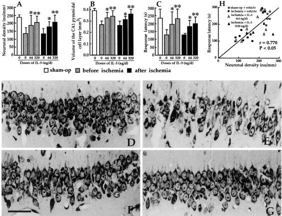
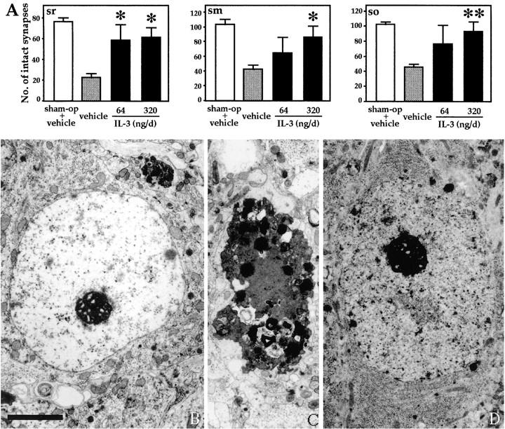
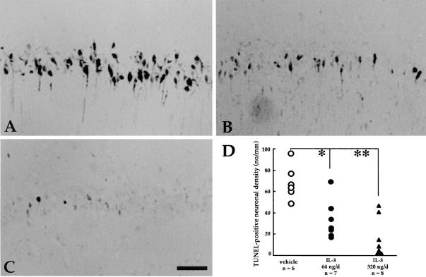
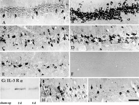
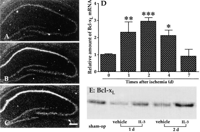
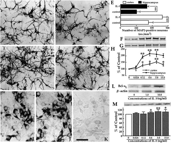
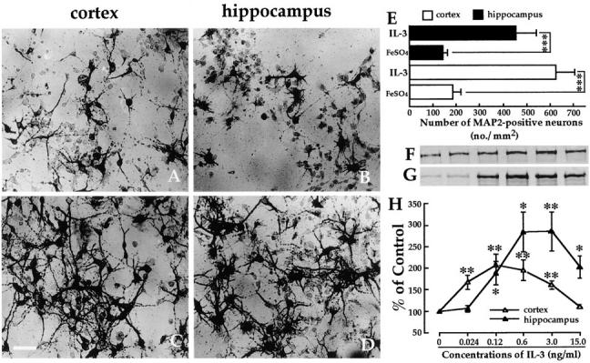
In the central nervous system, interleukin (IL)-3 has been shown to exert a trophic action only on septal cholinergic neurons in vitro and in vivo, but a widespread distribution of IL-3 receptor (IL-3R) in the brain does not conform to such a selective central action of the ligand. Moreover, the mechanism(s) underlying the neurotrophic action of IL-3 has not been elucidated, although an erythroleukemic cell line is known to enter apoptosis after IL-3 starvation possibly due to a rapid decrease in Bcl-2 expression. This in vivo study focused on whether IL-3 rescued noncholinergic hippocampal neurons from lethal ischemic damage by modulating the expression of Bcl-xL, a Bcl-2 family protein produced in the mature brain. 7-d IL-3 infusion into the lateral ventricle of gerbils with transient forebrain ischemia prevented significantly hippocampal CA1 neuron death and ischemia-induced learning disability. TUNEL (terminal deoxynucleotidyltransferase-mediated 2'-deoxyuridine 5'-triphosphate-biotin nick end labeling) staining revealed that IL-3 infusion caused a significant reduction in the number of CA1 neurons exhibiting DNA fragmentation 7 d after ischemia. The neuroprotective action of IL-3 appeared to be mediated by a postischemic transient upregulation of the IL-3R alpha subunit in the hippocampal CA1 field where IL-3Ralpha was barely detectable under normal conditions. In situ hybridization histochemistry and immunoblot analysis demonstrated that Bcl-xL mRNA expression, even though upregulated transiently in CA1 pyramidal neurons after ischemia, did not lead to the production of Bcl-xL protein in ischemic gerbils infused with vehicle. However, IL-3 infusion prevented the decrease in Bcl-xL protein expression in the CA1 field of ischemic gerbils. Subsequent in vitro experiments showed that IL-3 induced the expression of Bcl-xL mRNA and protein in cultured neurons with IL-3Ralpha and attenuated neuronal damage caused by a free radical-producing agent FeSO4. These findings suggest that IL-3 prevents delayed neuronal death in the hippocampal CA1 field through a receptor-mediated expression of Bcl-xL protein, which is known to facilitate neuron survival. Since IL-3Ralpha in the hippocampal CA1 region, even though upregulated in response to ischemic insult, is much less intensely expressed than that in the CA3 region tolerant to ischemia, the paucity of IL-3R interacting with the ligand may account for the vulnerability of CA1 neurons to ischemia.


Figures

Figure 1
Figure 1
(A and B) Effects of intracerebroventricular IL-3 infusion on CA1 neuronal density (A) and volume of the CA1 pyramidal cell layer (B) in ischemic gerbils. The infusion of IL-3 started 2 h before or just after 3-min ischemia, and continued for 7 d. Significant dose-dependent increases in CA1 neuronal density and volume of the CA1 pyramidal cell layer were noted in IL-3–infused ischemic gerbils compared with vehicle-infused ischemic animals. (C) Effect of IL-3 infusion on response latency in the passive avoidance task. The infusion of IL-3 significantly prolonged the response latency in a dose-dependent manner in ischemic gerbils compared with vehicle infusion. Each column in A–C represents mean ± SD (n = 6–8). *P <0.05, **P <0.01, Significantly different from the corresponding vehicle-infused ischemic group (statistical significance tested by the two-tailed Mann-Whitney U-test). (D–G) Photomicrographs of the hippocampal CA1 field: sham-operated animal infused with vehicle (D); ischemic animal infused with vehicle (E); ischemic animal infused with 64 ng/d of IL-3 (F); ischemic animal infused with 320 ng/d of IL-3 (G). Note that the infusion of IL-3, starting 2 h before 3-min ischemia, rescued a significant number of hippocampal CA1 pyramidal neurons. Sections were stained with 0.1% cresyl violet. Bar = 100 μm. (H) Positive correlation between the response latency in the passive avoidance task and the neuronal density of the hippocampal CA1 field, as evaluated by Pearson product–moment correlation analysis.
Figure 2
Figure 2
(A) Effect of IL-3 on the number of intact synapses in the three strata of the hippocampal CA1 region. The infusion of IL-3 or vehicle was started 2 h before 3-min ischemia, and continued for 7 d. Intact synapses in the strata radiatum (sr), moleculare (sm), and oriens (so) of the hippocampal CA1 region of ischemic gerbils with vehicle infusion were less numerous than in the strata of sham-operated (sham-op) animals, and the decrease in intact synapse number in the ischemic hippocampus was prevented significantly when IL-3 (64 or 320 ng/d) was infused into the lateral ventricle for 7 d. Each column represents mean ± SD (n = 6–8). *P <0.05, **P <0.01, Significantly different from the ischemic group with vehicle infusion (statistical significance tested by the two-tailed Mann-Whitney U-test). (B–D) Electron micrographs of the soma of ischemic hippocampal CA1 neurons treated with vehicle (B and C) or 320 ng/d of IL-3 (D). Note that the hippocampal CA1 pyramidal neurons in the vehicle- but not IL-3–treated ischemic gerbils were in the course of nuclear chromatin fragmentation and/or condensation (C), and that the cell nucleus of a vehicle-treated ischemic neuron at an early stage of degeneration (B) had an irregular euchromatin with electron density lower than that of an intact nuclear euchromatin (D). Bar = 5 μm.
Figure 3
Figure 3
(A–C) Photomicrographs of TUNEL-positive neurons in the hippocampal CA1 field of ischemic gerbils after 7-d infusion of vehicle or IL-3: vehicle (A); 64 ng/d of IL-3 (B); 320 ng/d of IL-3 (C). The infusion was started 2 h before 3-min ischemia, and continued for 7 d. Note that there were many TUNEL-positive cells in the hippocampal CA1 field of ischemic gerbils with vehicle infusion. Bar = 100 μm. (D) The number of TUNEL-positive neurons in the hippocampal CA1 field of ischemic gerbils. The number of TUNEL-positive neurons in the hippocampal CA1 field of IL-3 (64 or 320 ng/d)-infused ischemic gerbils was less than in vehicle-infused ischemic animals. *P <0.05; **P <0.01, Significantly different from the vehicle- infused ischemic group (statistical significance tested by the two-tailed Mann-Whitney U-test).
Figure 4
Figure 4
(A–F) Photomicrographs of IL-3Rα–immunoreactive neurons in the dorsal hippocampus: CA1 field of a sham-operated animal with vehicle infusion (A); CA3 field of a sham-operated animal with vehicle infusion (B); CA1 field of vehicle-treated animals 2 (C), 4 (D), and 7 (E) d after ischemia; CA1 field of a sham-operated animal stained with the immunoadsorbed primary antibody (F). Note a significant increase in IL-3Rα–immunoreactive neurons in the hippocampal CA1 field 2 (C) and 4 d (D) after ischemia. The CA3 field was more intensely labeled with the IL-3Rα antibody than was the CA1 field at any period examined. (G) Immunoblot analysis of IL-3Rα with a molecular mass of ∼70 kD in the CA1 field of sham-operated (sham-op) and ischemic gerbils. 40 μg of protein was loaded onto each lane. Ischemic insult appeared to increase IL-3Rα expression 2 and 4 d after ischemia. (H) CA1 field of an IL-3–infused animal 2 d after ischemia. (I) CA1 field of an IL-3–infused animal 4 d after ischemia. Note that IL-3 infusion caused an apparent decline in IL-3Rα–immunoreactive CA1 neurons 4 but not 2 d after ischemia. Bar = 100 μm.
Figure 5
Figure 5
(A–C) Dark-field photomicrographs showing Bcl-xL mRNA expression in the hippocampus of sham-operated and 3-min ischemic gerbils: hippocampus of a sham-operated animal (A); hippocampus 1 (B) and 2 (C) d after ischemia. Note a selective increase in Bcl-xL mRNA expression in the hippocampal CA1 field 1 and 2 d after ischemia. Bar = 200 μm. (D) Time course of Bcl-xL mRNA expression in the pyramidal cell layer of the CA1 field in 3-min ischemic gerbils. Relative amounts to the sham-operated control are expressed as mean ± SD. *P <0.05; **P < 0.01, ***P <0.001, Significantly different from the control (statistical significance tested by analysis of variance followed by Fisher's post hoc test). (E) Immunoblot analysis of Bcl-xL protein in the CA1 field of sham-operated (sham-op) and ischemic gerbils. Note a marked decline in Bcl-xL protein expression in the vehicle-treated CA1 field 1 d after ischemia, and that IL-3 treatment prevented the decrease in Bcl-xL protein expression in the CA1 field of ischemic gerbils 1 and 2 d after ischemia.
Figure 6
Figure 6
(A–D) Photomicrographs of MAP2-positive neurons in cultures: cortical (A) and hippocampal (B) neuron cultures without IL-3; cortical (C) and hippocampal (D) neuron cultures treated with 3.0 ng/ml of IL-3. Note that IL-3 significantly increased the number of MAP2-positive neurons. Bar = 200 μm. (E) Number of MAP2-positive neurons in cultures. The MAP2-positive neurons in the cultures treated with 3.0 ng/ml of IL-3 were more numerous than in the corresponding cultures without IL-3 (control). (F–H) MAP2 immunoblot analysis of IL-3–treated cortical (F) and hippocampal (G) neurons. Neurons cultured with 0–15.0 ng/ml of IL-3 were immunoblotted with an mAb against MAP2. MAP2-positive bands were observed in neurons without IL-3 treatment (lane 1 in F and G). MAP2-immunoreactive bands increased in intensity in cultures with 0.024–15.0 ng/ml of IL-3 (lanes 2–6 in F and G). Densitometric analysis of MAP2-immunoreactive bands showed that IL-3 enhanced the survival and possibly the neurite extension of cortical and hippocampal neurons in a dose-dependent manner (H). The data were obtained from four separate cultures and were expressed as a percentage of the corresponding control culture without IL-3. (I–K) Photomicrographs of IL-3Rα–immunoreactive neurons in cultures: cortical neuron culture (I), hippocampal neuron culture (J), and cortical neuron culture stained with the immunoadsorbed primary antibody (K). Note that IL-3Rα immunoreactions were abolished by adsorbing the primary antibody with the homologous antigen (K). Bar = 100 μm. (L) Semiquantitative RT-PCR analysis of Bcl-xL mRNA in cortical neurons cultured in the presence of 0, 1, or 10 ng/ml of IL-3. β-Actin was also amplified as an internal control from each sample. (M) Immunoblot analysis of Bcl-xL protein in cultured cortical neurons. IL-3 at concentrations of 0.6–15.0 ng/ml apparently induced Bcl-xL expression in the cultured neurons. The data were obtained from five separate cultures and were expressed as a percentage of the corresponding control culture. Each value in E, H, and M indicates mean ± SD. *P <0.05, **P <0.01, Significantly different from the corresponding control value (statistical significance tested by analysis of variance followed by Fisher's post hoc test).
Figure 7
Figure 7
(A–D) Photomicrographs of MAP2-positive neurons in cultures exposed to FeSO4: cortical (A) and hippocampal (B) neurons in cultures treated only with FeSO4; cortical (C) and hippocampal (D) neurons in cultures pretreated with 3.0 ng/ml of IL-3 for 3 d, then treated with FeSO4. Note that IL-3 treatment significantly increased the number of MAP2-positive neurons. Bar = 200 μm. (E) Number of MAP2-positive neurons in cultures exposed to FeSO4. The MAP2-positive neurons in the cultures treated with 3.0 ng/ml of IL-3 were more numerous than in the corresponding control cultures without IL-3 (FeSO4). (F–H) MAP2 immunoblot analysis of IL-3–treated cortical (F) and hippocampal (G) neurons exposed to FeSO4. Samples cultured for 3 d with 0–15.0 ng/ml of IL-3 and then treated with FeSO4 were immunoblotted with an mAb against MAP2. Neurons cultured with FeSO4 but without IL-3 pretreatment showed extremely weak immunoreactive bands (lane 1 in F and G). In neurons treated with 0.12–3.0 ng/ml of IL-3 and then with FeSO4, the intensity of MAP2-immunoreactive bands significantly increased even if FeSO4 existed in the cultures (lanes 3–5 in F and G). The densitometric analysis of MAP2-immunoreactive bands showed that IL-3 protected the cultured neurons against FeSO4-induced oxidative injuries in a dose-dependent manner (H). The data were obtained from four separate cultures and were expressed as a percentage of the corresponding control culture. Each value in E and H indicates mean ± SD. *P <0.05, **P <0.01, ***P < 0.001, Significantly different from the corresponding control value (statistical significance tested by analysis of variance followed by Fisher's post hoc test).

Similar articles

Cited by

References

    1. Schrader JW. The pan specific hemopoietin of activated T-lymphocytes (interleukin-3) Annu Rev Immunol. 1986;4:205–230. - PubMed
    1. Frendl G, Beller DI. Regulation of macrophage activation by IL-3. J Immunol. 1990;144:3392–3399. - PubMed
    1. Frendl G. Interleukin-3: from colony-stimulating factor to pluripotent immunoregulatory cytokine. J Immunol. 1992;14:421–430. - PubMed
    1. Ihle JN, Weinstein Y. Immunological regulation of hematopoietic/lymphoid stem cell differentiation by interleukin 3. Adv Immunol. 1986;39:1–50. - PubMed
    1. Clarke SC, Kamen R. The human hematopoi-etic colony-stimulating factors. Science. 1987;236:1229–1237. - PubMed

Publication types

MeSH terms