

Early function of Pax5 (BSAP) before the pre-B cell receptor stage of B lymphopoiesis
- PMID: 9705955
- PMCID: PMC2213350
- DOI: 10.1084/jem.188.4.735
Early function of Pax5 (BSAP) before the pre-B cell receptor stage of B lymphopoiesis
Abstract
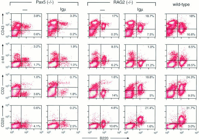
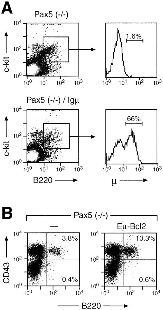
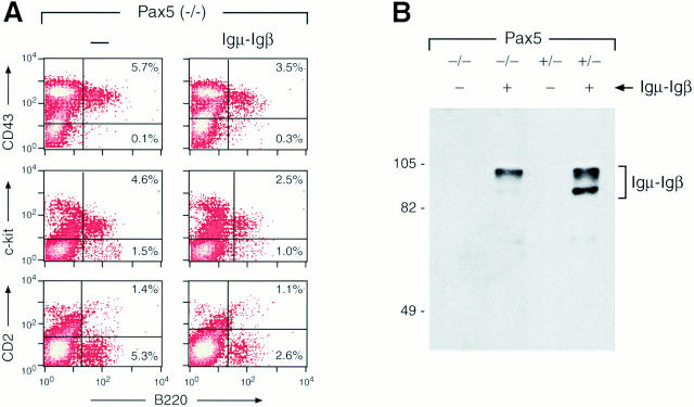
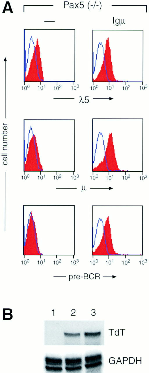
The formation of the pre-B cell receptor (BCR) corresponds to an important checkpoint in B cell development that selects pro-B (pre-BI) cells expressing a functionally rearranged immunoglobulin mu (Igmu) heavy chain protein to undergo the transition to the pre-B (pre-BII) cell stage. The pre-BCR contains, in addition to Igmu, the surrogate light chains lambda5 and VpreB and the signal transducing proteins Igalpha and Igbeta. The absence of one of these pre-BCR components is known to arrest B cell development at the pre-BI cell stage. Disruption of the Pax5 gene, which codes for the B cell-specific activator protein (BSAP), also blocks adult B lymphopoiesis at the pre-BI cell stage. Moreover, expression of the mb-1 (Igalpha) gene and VH-to-DHJH recombination at the IgH locus are reduced in Pax5-deficient B lymphocytes approximately 10- and approximately 50-fold, respectively. Here we demonstrate that complementation of these deficiencies in pre-BCR components by expression of functionally rearranged Ig mu and chimeric Igmu-Igbeta transgenes fails to advance B cell development to the pre-BII cell stage in Pax5 (-/-) mice in contrast to RAG2 (-/-) mice. Furthermore, the pre-BCR is stably expressed on cultured pre-BI cells from Igmu transgenic, Pax5-deficient bone marrow, but is unable to elicit its normal signaling responses. In addition, the early developmental block is unlikely to be caused by the absence of a survival signal, as it could not be rescued by expression of a bcl2 transgene in Pax5-deficient pre-BI cells. Together, these data demonstrate that the absence of Pax5 arrests adult B lymphopoiesis at an early developmental stage that is unresponsive to pre-BCR signaling.
Figures
Similar articles
-
Essential functions of Pax5 (BSAP) in pro-B cell development: difference between fetal and adult B lymphopoiesis and reduced V-to-DJ recombination at the IgH locus.Genes Dev. 1997 Feb 15;11(4):476-91. doi: 10.1101/gad.11.4.476. Genes Dev. 1997. PMID: 9042861
-
Long-term in vivo reconstitution of T-cell development by Pax5-deficient B-cell progenitors.Nature. 1999 Oct 7;401(6753):603-6. doi: 10.1038/44164. Nature. 1999. PMID: 10524629
-
Control of pre-BCR signaling by Pax5-dependent activation of the BLNK gene.Immunity. 2002 Oct;17(4):473-85. doi: 10.1016/s1074-7613(02)00418-1. Immunity. 2002. PMID: 12387741
-
Essential functions of Pax-5 (BSAP) in pro-B cell development.Immunobiology. 1997 Dec;198(1-3):227-35. doi: 10.1016/S0171-2985(97)80043-5. Immunobiology. 1997. PMID: 9442394 Review.
-
Loss- and gain-of-function mutations reveal an important role of BSAP (Pax-5) at the start and end of B cell differentiation.Semin Immunol. 1998 Apr;10(2):133-42. doi: 10.1006/smim.1998.0115. Semin Immunol. 1998. PMID: 9618759 Review.
Cited by
-
Rack1 regulates B-cell development and function by binding to and stabilizing the transcription factor Pax5.Cell Mol Immunol. 2024 Nov;21(11):1282-1295. doi: 10.1038/s41423-024-01213-2. Epub 2024 Sep 10. Cell Mol Immunol. 2024. PMID: 39256480
-
The BCR-ABL1 kinase bypasses selection for the expression of a pre-B cell receptor in pre-B acute lymphoblastic leukemia cells.J Exp Med. 2004 Mar 1;199(5):673-85. doi: 10.1084/jem.20031637. J Exp Med. 2004. PMID: 14993251 Free PMC article.
-
Immunophenotyping of Murine Precursor B-Cell Leukemia/Lymphoma: A Comparison of Immunohistochemistry and Flow Cytometry.Vet Pathol. 2019 Nov;56(6):950-958. doi: 10.1177/0300985819852138. Epub 2019 Jun 6. Vet Pathol. 2019. PMID: 31170889 Free PMC article.
-
Sox4 is required for the survival of pro-B cells.J Immunol. 2013 Mar 1;190(5):2080-9. doi: 10.4049/jimmunol.1202736. Epub 2013 Jan 23. J Immunol. 2013. PMID: 23345330 Free PMC article.
-
Pax5 is required for recombination of transcribed, acetylated, 5' IgH V gene segments.Genes Dev. 2003 Jan 1;17(1):37-42. doi: 10.1101/gad.1031403. Genes Dev. 2003. PMID: 12514097 Free PMC article.
References
-
- Rajewsky K. Clonal selection and learning in the antibody system. Nature. 1996;381:751–758. - PubMed
-
- Borst J, Jacobs H, Brouns G. Composition and function of T-cell receptor and B-cell receptor complexes on precursor lymphocytes. Curr Opin Immunol. 1996;8:181–190. - PubMed
-
- Kitamura D, Roes J, Kühn R, Rajewsky K. A B cell–deficient mouse by targeted disruption of the membrane exon of the immunoglobulin μ chain gene. Nature. 1991;350:423–426. - PubMed
-
- Kitamura D, Kudo A, Schaal S, Müller W, Melchers F, Rajewsky K. A critical role of λ5 protein in B cell development. Cell. 1992;69:823–831. - PubMed
-
- Gong S, Nussenzweig MC. Regulation of an early developmental checkpoint in the B cell pathway by Igβ. Science. 1996;272:411–414. - PubMed
Publication types
MeSH terms
Substances
LinkOut - more resources
Full Text Sources
Medical
Molecular Biology Databases
Research Materials
Miscellaneous
