

Complementation of lymphotoxin alpha knockout mice with tumor necrosis factor-expressing transgenes rectifies defective splenic structure and function
- PMID: 9705956
- PMCID: PMC2213356
- DOI: 10.1084/jem.188.4.745
Complementation of lymphotoxin alpha knockout mice with tumor necrosis factor-expressing transgenes rectifies defective splenic structure and function
Abstract
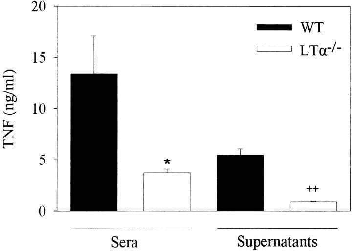
Lymphotoxin (LT)alpha knockout mice, as well as double LTalpha/tumor necrosis factor (TNF) knockout mice, show a severe splenic disorganization with nonsegregating T/B cell zones and complete absence of primary B cell follicles, follicular dendritic cell (FDC) networks, and germinal centers. In contrast, as shown previously and confirmed in this study, LTbeta-deficient mice show much more conserved T/B cell areas and a reduced but preserved capacity to form germinal centers and FDC networks. We show here that similar to the splenic phenotype of LTbeta-deficient mice, complementation of LTalpha knockout mice with TNF-expressing transgenes leads to a p55 TNF receptor-dependent restoration of B/T cell zone segregation and a partial preservation of primary B cell follicles, FDC networks, and germinal centers. Notably, upon lipopolysaccharide challenge, LTalpha knockout mice fail to produce physiological levels of TNF both in peritoneal macrophage supernatants and in their serum, indicating a coinciding deficiency in TNF expression. These findings suggest that defective TNF expression contributes to the complex phenotype of the LTalpha knockout mice, and uncover a predominant role for TNF and its p55 TNF receptor in supporting, even in the absence of LTalpha, the development and maintenance of splenic B cell follicles, FDC networks, and germinal centers.
Figures


Similar articles
-
The complementation of lymphotoxin deficiency with LIGHT, a newly discovered TNF family member, for the restoration of secondary lymphoid structure and function.Eur J Immunol. 2002 Jul;32(7):1969-79. doi: 10.1002/1521-4141(200207)32:7<1969::AID-IMMU1969>3.0.CO;2-M. Eur J Immunol. 2002. PMID: 12115617
-
Tumor necrosis factor and the p55TNF receptor are required for optimal development of the marginal sinus and for migration of follicular dendritic cell precursors into splenic follicles.Cell Immunol. 2000 Apr 10;201(1):33-41. doi: 10.1006/cimm.2000.1636. Cell Immunol. 2000. PMID: 10805971
-
Distinct contributions of TNF and LT cytokines to the development of dendritic cells in vitro and their recruitment in vivo.Blood. 2003 Feb 15;101(4):1477-83. doi: 10.1182/blood.V101.4.1477. Blood. 2003. PMID: 12560241
-
Lymphotoxin-alpha-deficient and TNF receptor-I-deficient mice define developmental and functional characteristics of germinal centers.Immunol Rev. 1997 Apr;156:137-44. doi: 10.1111/j.1600-065x.1997.tb00965.x. Immunol Rev. 1997. PMID: 9176705 Review.
-
Peyer's patch organogenesis--cytokines rule, OK?Gut. 1997 Nov;41(5):707-9. doi: 10.1136/gut.41.5.707. Gut. 1997. PMID: 9414984 Free PMC article. Review.
Cited by
-
Control of experimental Trypanosoma brucei infections occurs independently of lymphotoxin-alpha induction.Infect Immun. 2002 Mar;70(3):1342-51. doi: 10.1128/IAI.70.3.1342-1351.2002. Infect Immun. 2002. PMID: 11854219 Free PMC article.
-
Tumor-Associated Tertiary Lymphoid Structures: Gene-Expression Profiling and Their Bioengineering.Front Immunol. 2017 Jun 30;8:767. doi: 10.3389/fimmu.2017.00767. eCollection 2017. Front Immunol. 2017. PMID: 28713385 Free PMC article.
-
Mature follicular dendritic cell networks depend on expression of lymphotoxin beta receptor by radioresistant stromal cells and of lymphotoxin beta and tumor necrosis factor by B cells.J Exp Med. 1999 Jan 4;189(1):159-68. doi: 10.1084/jem.189.1.159. J Exp Med. 1999. PMID: 9874572 Free PMC article.
-
TNFα-dependent development of lymphoid tissue in the absence of RORγt⁺ lymphoid tissue inducer cells.Mucosal Immunol. 2014 May;7(3):602-14. doi: 10.1038/mi.2013.79. Epub 2013 Oct 16. Mucosal Immunol. 2014. PMID: 24129162 Free PMC article.
-
Role of tumour necrosis factor alpha in experimental arthritis: separate activity of interleukin 1beta in chronicity and cartilage destruction.Ann Rheum Dis. 1999 Nov;58 Suppl 1(Suppl 1):I40-8. doi: 10.1136/ard.58.2008.i40. Ann Rheum Dis. 1999. PMID: 10577972 Free PMC article. Review.
References
-
- Browning JL, Ngam-ek A, Lawton P, DeMarinis J, Tizard R, Chow EP, Hession C, O'Brine-Greco B, Foley SF, Ware CF. Lymphotoxin beta, a novel member of the TNF family that forms a heteromeric complex with lymphotoxin on the cell surface. Cell. 1993;72:847–856. - PubMed
-
- Kriegler M, Perez C, DeFay K, Albert I, Lu SD. A novel form of TNF/cachectin is a cell surface cytotoxic transmembrane protein: ramifications for the complex physiology of TNF. Cell. 1988;53:45–53. - PubMed
-
- Vandenabeele P, Declercq W, Beyaert R, Fiers W. Two tumour necrosis factor receptors: structure and function. Trends Cell Biol. 1995;5:392–399. - PubMed
-
- Crowe PD, VanArsdale TL, Walter BN, Ware CF, Hession C, Ehrenfels B, Browning JL, Din WS, Goodwin RG, Smith CA. A lymphotoxin-beta-specific receptor. Science. 1994;264:707–710. - PubMed
Publication types
MeSH terms
Substances
LinkOut - more resources
Full Text Sources
Other Literature Sources
Molecular Biology Databases
