. 1998 Aug 17;188(4):765-72.
doi: 10.1084/jem.188.4.765.

Antigen receptor engagement turns off the V(D)J recombination machinery in human tonsil B cells

Affiliations

Antigen receptor engagement turns off the V(D)J recombination machinery in human tonsil B cells

E Meffre et al. J Exp Med. .

Abstract

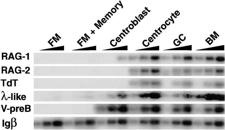
The germinal center (GC) is an anatomic compartment found in peripheral lymphoid organs, wherein B cells undergo clonal expansion, somatic mutation, switch recombination, and reactivate immunoglobulin gene V(D)J recombination. As a result of somatic mutation, some GC B cells develop higher affinity antibodies, whereas others suffer mutations that decrease affinity, and still others may become self-reactive. It has been proposed that secondary V(D)J rearrangements in GCs might rescue B cells whose receptors are damaged by somatic mutations. Here we present evidence that mature human tonsil B cells coexpress conventional light chains and recombination associated genes, and that they extinguish recombination activating gene and terminal deoxynucleotidyl transferase expression when their receptors are cross-linked. Thus, the response of the recombinase to receptor engagement in peripheral B cells is the opposite of the response in developing B cells to the same stimulus. These observations suggest that receptor revision is a mechanism for receptor diversification that is turned off when antigen receptors are cross-linked by the cognate antigen.


Figures

Figure 1
Figure 1
Ligation-mediated (LM)-PCR assay for detecting signal breaks in human GC B cells. (a) Scheme for LM-PCR strategy to detect RSS DNA breaks within the human Ig κ light chain locus. A primary VκJκ1 rearrangement is represented at the κ locus, and after V(D)J recombinase activation, downstream Jκ segments may be chosen for a secondary rearrangement; Jκ5 is depicted. Blunt signal ends at the nonamer–heptamer sequences (triangles) are ligated to BW linkers. Linker-ligated DNA molecules are then amplified by PCR using two sets of specific primers (see Materials and Methods). (b) Jκ signal breaks in human GC B cells. LM-PCR for JH6 (top) and Jκ5 (middle) signal breaks in human mononuclear bone marrow (BM) cells, naive CD38IgD+ FM tonsil B cells, and CD38+IgD GC B cells (left) and in centroblasts CD38+CD77+ and centrocytes CD38+CD77 (right). PCR products were visualized with locus-specific probes. PCR from a region overlapping the germline Jκ5 segment was used as a control for DNA loading (bottom).
Figure 1
Figure 1
Ligation-mediated (LM)-PCR assay for detecting signal breaks in human GC B cells. (a) Scheme for LM-PCR strategy to detect RSS DNA breaks within the human Ig κ light chain locus. A primary VκJκ1 rearrangement is represented at the κ locus, and after V(D)J recombinase activation, downstream Jκ segments may be chosen for a secondary rearrangement; Jκ5 is depicted. Blunt signal ends at the nonamer–heptamer sequences (triangles) are ligated to BW linkers. Linker-ligated DNA molecules are then amplified by PCR using two sets of specific primers (see Materials and Methods). (b) Jκ signal breaks in human GC B cells. LM-PCR for JH6 (top) and Jκ5 (middle) signal breaks in human mononuclear bone marrow (BM) cells, naive CD38IgD+ FM tonsil B cells, and CD38+IgD GC B cells (left) and in centroblasts CD38+CD77+ and centrocytes CD38+CD77 (right). PCR products were visualized with locus-specific probes. PCR from a region overlapping the germline Jκ5 segment was used as a control for DNA loading (bottom).
Figure 2
Figure 2
RAG, TdT, λ-like, and V-preB gene expression in human tonsil B cell subsets. RNA from FACS®-sorted CD38IgD+ FM B lymphocytes, CD38CD77 FM and memory B cells, CD38+CD77+ GC centroblasts, CD38+CD77 GC centrocytes, CD38+IgD total GC B cells, and mononuclear bone marrow (BM) cells was analyzed by semiquantitative RT-PCR and visualized with labeled oligonucleotide probes. Igβ RT-PCR was used as a B cell–specific mRNA loading control. PCR assays were designed to distinguish between genomic DNA and mRNA using primers on different exons. PhosphorImager analysis showed that there is a fivefold difference in the amount of RAG1 mRNA amplified between centroblasts and centrocytes.
Figure 3
Figure 3
ΨL cell surface expression on GC B cells. (a) Flow cytometric analysis were performed on human tonsil B cells using anti-CD38, anti–V-preB, anti-Igκ, or anti-Igλ. Two different patient samples are shown. (b) Flow cytometric analysis of immunized wild-type and λ5-targeted (λ5−/−) mouse spleen cells using anti-B220, anti-GL7, anti-κ, and anti-λ, and either LM34 or SL156 anti-λ5 antibodies (42).
Figure 3
Figure 3
ΨL cell surface expression on GC B cells. (a) Flow cytometric analysis were performed on human tonsil B cells using anti-CD38, anti–V-preB, anti-Igκ, or anti-Igλ. Two different patient samples are shown. (b) Flow cytometric analysis of immunized wild-type and λ5-targeted (λ5−/−) mouse spleen cells using anti-B220, anti-GL7, anti-κ, and anti-λ, and either LM34 or SL156 anti-λ5 antibodies (42).
Figure 4
Figure 4
ΨL expression in human tonsil B cells. Immunohistochemical staining of human tonsil sections was performed using anti–V-preB (left), anti-CD21 (middle), or both (right). GCs, FM, and the T cell zones (T zone) are indicated.
Figure 5
Figure 5
Regulated expression of RAG, TdT, λ-like, and V-preB genes. (a) Human tonsil CD38IgD+ FM or CD38+IgD GC B cells were sorted (d0) and cultured on CD40L-transfected fibroblasts with or without anti-κ+λ (α Ig) at 10 μg/ml, or IL-2, IL-4, or IL-10 for 3 d (d3). Gene expression was analyzed by RT-PCR as described above. (b) CD38+IgD GC B cells were cultured with CD40L alone or with CD40L and an irrelevant IgG control (Control Ig), or with intact anti-κ (Ig α–κ) or anti-λ (Ig α–λ) or both (Ig α–κ + α–λ) or with Fab′2 anti-κ or anti-λ (Fab′2 α–κ + α–λ). Antibody concentrations are indicated in micrograms per milliliter. (c) GC cells reanalyzed by flow cytometry using anti-CD38 and anti–V-preB mAbs after 3 d of culture with CD40L and the indicated antibodies. GC B cells cultured with Ig α–κ + α–λ or Fab′2 α–κ + α–λ antibodies contained 1.5–2-fold more B cells than those with cultured with control antibody, and the percentage of dead cells was 40–50% in all of cultures as determined by trypan blue exclusion.
Figure 5
Figure 5
Regulated expression of RAG, TdT, λ-like, and V-preB genes. (a) Human tonsil CD38IgD+ FM or CD38+IgD GC B cells were sorted (d0) and cultured on CD40L-transfected fibroblasts with or without anti-κ+λ (α Ig) at 10 μg/ml, or IL-2, IL-4, or IL-10 for 3 d (d3). Gene expression was analyzed by RT-PCR as described above. (b) CD38+IgD GC B cells were cultured with CD40L alone or with CD40L and an irrelevant IgG control (Control Ig), or with intact anti-κ (Ig α–κ) or anti-λ (Ig α–λ) or both (Ig α–κ + α–λ) or with Fab′2 anti-κ or anti-λ (Fab′2 α–κ + α–λ). Antibody concentrations are indicated in micrograms per milliliter. (c) GC cells reanalyzed by flow cytometry using anti-CD38 and anti–V-preB mAbs after 3 d of culture with CD40L and the indicated antibodies. GC B cells cultured with Ig α–κ + α–λ or Fab′2 α–κ + α–λ antibodies contained 1.5–2-fold more B cells than those with cultured with control antibody, and the percentage of dead cells was 40–50% in all of cultures as determined by trypan blue exclusion.

Similar articles

Cited by

References

    1. Tonegawa S. Somatic generation of antibody diversity. Nature. 1983;302:575–581. - PubMed
    1. Willerford DM, Swat W, Alt FW. Developmental regulation of V(D)J recombination and lymphocyte differentiation. Curr Opin Genet Dev. 1996;6:603–609. - PubMed
    1. Schatz DG, Oettinger MA, Baltimore D. The V(D)J recombination activating gene, RAG-1. Cell. 1989;59:1035–1048. - PubMed
    1. Oettinger MA, Schatz DG, Gorka C, Baltimore D. RAG-1 and RAG-2, adjacent genes that synergistically activate V(D)J recombination. Science. 1990;248:1517–1523. - PubMed
    1. Shinkai Y, Rathbun G, Lam K-P, Oltz EM, Stewart V, Mendelsohn M, Charron J, Datta M, Young F, Stall AM, Alt FW. RAG-2 deficient mice lack mature lymphocytes owing to inability to initiate V(D)J rearrangement. Cell. 1992;68:855–867. - PubMed

Publication types

MeSH terms