. 1998 Jul 15;510 ( Pt 2)(Pt 2):333-45.
doi: 10.1111/j.1469-7793.1998.333bk.x.

The sulphonylurea receptor SUR1 regulates ATP-sensitive mouse Kir6.2 K+ channels linked to the green fluorescent protein in human embryonic kidney cells (HEK 293)

Affiliations

The sulphonylurea receptor SUR1 regulates ATP-sensitive mouse Kir6.2 K+ channels linked to the green fluorescent protein in human embryonic kidney cells (HEK 293)

S A John et al. J Physiol. .

Abstract

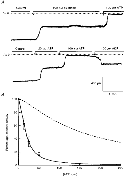
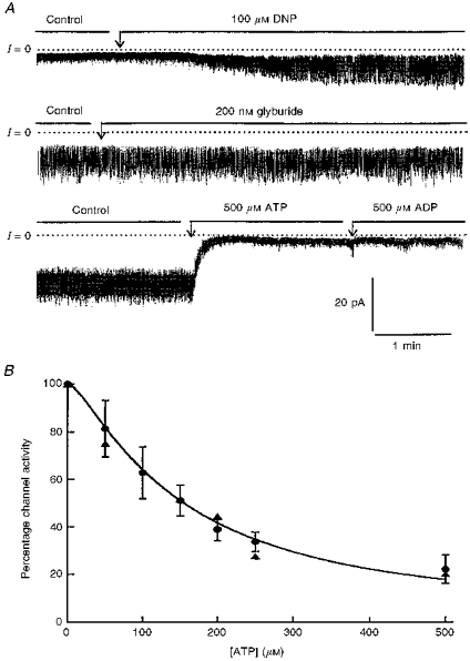
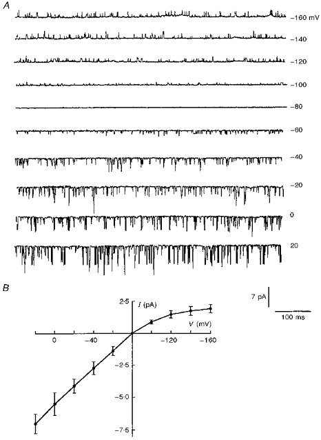
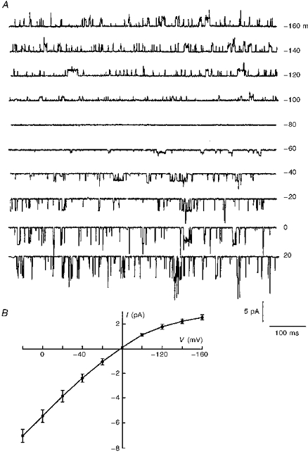
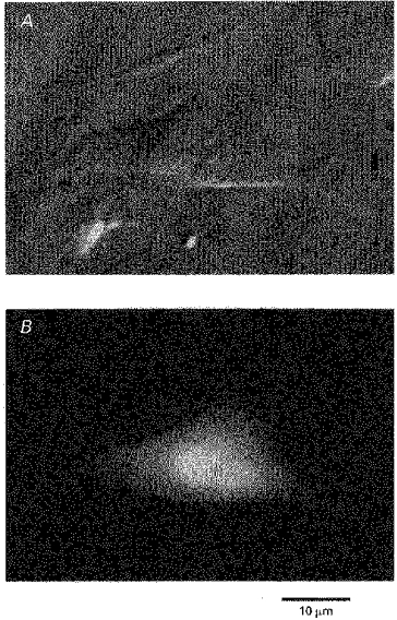
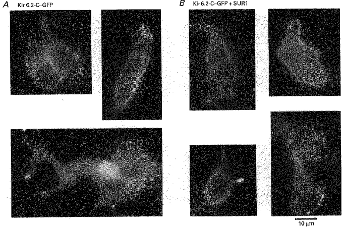
1. Using a chimeric protein comprising the green fluorescent protein (GFP) linked to the C-terminus of the K+ channel protein mouse Kir6.2 (Kir6.2-C-GFP), the interactions between the sulphonylurea receptor SUR1 and Kir6.2 were investigated in transfected human embryonic kidney cells (HEK 293) by combined imaging and patch clamp techniques. 2. HEK 293 cells transfected with mouse Kir6.2-C-GFP and wild-type Kir6.2 exhibited functional K+ channels independently of SUR1. These channels were inhibited by ATP (IC50 = 150 microM), but were not responsive to stimulation by ADP or inhibition by sulphonylureas. Typically 15 +/- 7 active channels were found in an excised patch. 3. The distribution of Kir6.2-C-GFP protein was investigated by imaging of GFP fluorescence. There was a lamellar pattern of fluorescence labelling inside the cytoplasm (presumably associated with the endoplasmic reticulum and the Golgi apparatus) and intense punctate labelling near the cell membrane, but little fluorescence was associated with the plasma membrane. 4. In contrast, cells co-transfected with Kir6.2-C-GFP and SUR1 exhibited intense uniform plasma membrane labelling, and the lamellar and punctate labelling seen without SUR1 was no longer prominent. 5. In cells co-transfected with Kir6.2-C-GFP and SUR1, strong membrane labelling was associated with very high channel activity, with 484 +/- 311 active channels per excised patch. These K+ channels were sensitive to inhibition by ATP (IC50 = 17 microM), stimulated by ADP and inhibited by sulphonylureas. 6. We conclude that co-expression of SUR1 and Kir6.2 generates channels with the properties of native KATP channels. In addition, SUR1 promotes uniform insertion of Kir6.2-C-GFP into the plasma membrane and a 35-fold increase in channel activity, suggesting that SUR1 facilitates protein trafficking of Kir6.2 into the plasma membrane.


Figures

Figure 7
Figure 7. Modulation by adenine nucleotides and sulphonylurea of K+ channels generated by Kir6.2-C–GFP in the presence of SUR1
A, current recordings obtained in excised inside-out patches (upper and lower traces). Both recordings are from the same patch, but the data in the lower trace were obtained first. The membrane holding potential was −10 mV. The currents are inward currents and channels open downward. The dotted line represents zero current when all channels are closed. The upper trace demonstrates partial channel inhibition by glyburide and almost complete block by 100 μM ATP. The lower trace illustrates the dose-dependent inhibition of Kir6.2 K+ channels by ATP and the reversal of inhibition by ADP. B, ATP-dependent inhibition of Kir6.2-C–GFP + SUR1 (•). Data points are averages of four measurements obtained from four different patches at various ATP concentrations. Channel activity was normalized to the activity measured in the same patch in the absence of nucleotide. The individual values were then averaged, plotted as a function of ATP, and fitted with the equation given in the legend to Fig. 4. The fit of the data represented by the continuous line yielded K = 17.2 mM and n = 1.56. The dotted line which is shown for comparison represents the sensitivity to ATP of Kir6.2 in the absence of SUR1 (see Fig. 4).
Figure 4
Figure 4. Modulation of Kir6.2-C–GFP by adenine nucleotides and sulphonylureas
A, current recordings obtained in the cell-attached mode (upper trace) and with excised inside-out patches (middle and lower traces). The membrane holding potential was −25 mV for the upper trace and −20 mV for the middle and lower traces. In all cases currents are inward and channels are shown to open downward. The dotted line represents zero current when all channels are closed. The upper trace illustrates the effect of the mitochondrial inhibitor dinitrophenol (DNP). Within 2 min, DNP stimulated channel activity, and the effect reached a maximum after about 3 min. The stimulatory effect of DNP was poorly reversible (not shown). The middle trace illustrates the lack of channel inhibition by the sulphonylurea glyburide. The lower trace represents the inhibitory effect of ATP and the lack of channel stimulation by MgADP. B, ATP-dependent inhibition of Kir6.2-C–GFP (•) and Kir6.2 +β-C-GFP (▴). Data points are averages of six measurements (•) and two measurements (▴) obtained from ten patches, with various ATP concentrations. To allow for comparison between patches, the channel activity was normalized to the activity measured in the same patch in the absence of nucleotide. The individual values were then averaged, plotted as a function of ATP, and fitted with the equation: I/Io = 100/(1 + (C/K)n), where C is the ATP concentration, K is the IC50 constant and n is a Hill coefficient. The fit of the data represented by the continuous line yielded K = 151 mM and n = 1.3.
Figure 3
Figure 3. Effects of membrane potential on single channel currents generated by Kir6.2-C–GFP
A, current recordings measured at varying holding potentials in the cell-attached patch configuration, with 5 mM K+ in the bath. Downward deflections represent inward currents. B, the I-V relationship obtained from recordings similar to those in A. The values on the horizontal axis and on the right side of A are pipette holding potentials.
Figure 6
Figure 6. Effects of membrane potential on single channel currents generated by Kir6.2-C–GFP + SUR1
A, current recordings obtained at varying holding potentials in the cell-attached patch configuration, in the presence of 5 mM K+ in the bath. Downward deflections represent inward currents. B, the I-V relationship obtained from recordings similar to those shown in A. The values on the horizontal axis and on the right margin of A are pipette holding potentials.
Figure 1
Figure 1. Expression of GFP and Kir6.2 in HEK 293 cells
Co-transfection with plasmids encoding GFP and the ATP-dependent K+ channel Kir6.2 yielded cells with green fluorescence labelling (B). The fluorescence was evenly distributed throughout the cytoplasm. Comparison of the DIC image in A with the fluorescence image in B indicates that only some of the cells were transfected with GFP.
Figure 2
Figure 2. Expression of the chimeric Kir6.2-C–GFP, alone (A) and with SUR1 (B)
A, three images, each obtained from a different transfection experiment representative of the distribution of GFP linked to Kir6.2 in HEK 293 cells. Bright, punctate fluorescent structures, possibly associated with vesicles, were found near the cell membrane. Strong fluorescence in a lamellar pattern was also found and tentatively identified as the endoplasmic reticulum and/or the Golgi apparatus. Dim fluorescence was associated with the plasma membrane. B, four images, which are from four different transfection experiments, representative of cells co-transfected with Kir6.2-C–GFP and SUR1, illustrating the effect of SUR1 on Kir6.2-C–GFP localization. Bright, uniform fluorescence is now present over the plasma membrane. In contrast with the images from cells transfected with Kir6.2-C–GFP alone, the lamellar and punctate labelling patterns are no longer observed, suggesting that Kir6.2-C–GFP is now uniformly incorporated in the plasma membrane. For further information on cellular distribution of GFP, including 3-D images, see the Web site: http://www.medsch.ucla.edu/som/physio/faculty/br.htm
Figure 5
Figure 5. Kinetic behaviour of wild-type Kir6.2 and modulation by adenine nucleotides
The upper trace is a recording obtained in an excised inside-out patch of a cell transfected with wild-type Kir6.2, and β-C-GFP as a marker. These data illustrate the inhibitory effect of ATP and the lack of channel stimulation by MgADP. The lower trace is a representative segment of the upper one, displayed using an expanded time scale to illustrate the short open times of wild-type Kir6.2. The labels to the left of this lower trace indicate when all the channels are closed (C), when one channel is open (1) and when two channels are open (2). In this experiment the membrane holding potential was −55 mV.

References

    1. Aguilar-Bryan L, Nichols CG, Wechsler SW, Clement JP, IV, Boyd AE, III, Gonzalez G, Herrera-Sosa H, Nguy K, Bryan J, Nelson DA. Cloning of the β cell high-affinity sulfonylurea receptor: A regulator of insulin secretion. Science. 1995;268:423–426. - PubMed
    1. Ashcroft FM. Adenosine 5′-triphosphate-sensitive potassium channels. Annual Review of Neuroscience. 1988;11:97–118. 10.1146/annurev.ne.11.030188.000525. - DOI - PubMed
    1. Chalfie M, Tu Y, Euskirchen G, Ward WW, Prasher DC. Green fluorescent protein as a marker for gene expression. Science. 1994;263:802–805. - PubMed
    1. Cockett MI, Ochalski R, Benwell K, Franco R, Wardell-Swanson J. Simultaneous expression of multi-subunit proteins in mammalian cells using a convenient set of mammalian cell expression vectors. Biotechniques. 1977;23:402–405. - PubMed
    1. Daut J, Maier-Rudolph W, Von Beckerath N, Mehrke G, Gunther K. Hypoxic dilation of coronary arteries is mediated by ATP-sensitive potassium channels. Science. 1990;247:1341–1344. - PubMed

Publication types

MeSH terms

LinkOut - more resources