. 1998 Jul 15;510 ( Pt 2)(Pt 2):421-7.
doi: 10.1111/j.1469-7793.1998.421bk.x.

Muscarinic IPSPs in rat striatal cholinergic interneurones

Affiliations

Muscarinic IPSPs in rat striatal cholinergic interneurones

P Calabresi et al. J Physiol. .

Abstract

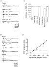
1. Intracellular recordings were made from neurones in slice of rat striatum in vitro. 2. The forty-nine neurones studied were immunoreactive for choline acetyltransferase and had the electrophysiological characteristics typical of large aspiny interneurones. 3. Focal stimulation of the slice elicited a hyperpolarizing inhibitory postsynaptic potential in thirty-five neurones. This IPSP lasted 0.5-1 s and reversed polarity at a membrane potential which was dependent on the logarithm of the extracellular potassium concentration. 4. The IPSP was reversibly blocked by scopolamine and methoctramine, which has some selectivity for M2 subtype of muscarinic receptor. It was unaffected by 6-cyano-7-nitroquinoxaline-2,3-dione (10 microM), DL-2-amino-phosphonovaleric acid (30 microM) and bicuculline (30 microM). 5. Exogenous acetylcholine and muscarine also hyperpolarized the neurones, and this was blocked by methoctramine by not by pirenzepine, which is an M1 receptor-selective antagonist. 6. The findings demonstrate that muscarinic IPSPs occur in the central nervous system. The IPSP may mediate an 'autoinhibition' of striatal cholinergic neurone activity.


Figures
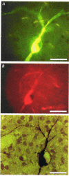
Figure 1
Figure 1. Identification of cholinergic interneurones after intracellular recording
A, the cell was injected with biocytin and visualized by fluorescein isothiocyanate-conjugated avidin. Note the large body and the roundish unstained nucleus. B, choline acetyltransferase immunoreactivity visualized by tetramethylrhodamine isothiocyanate-conjugated secondary antibody. C, permanent staining after incubation with avidin-biotin-peroxidase complex and diaminobenzidine. The three photomicrographs show the same cell. Note the beaded aspinous dendrites. Scale bar, 25 μm.
Figure 2
Figure 2. Cholinergic IPSPs in striatal interneurones
A, a single pulse stimulus (arrows) evoked a fast depolarizing synaptic potential followed by a slower hyperpolarizing synaptic potential (IPSP). The depolarization was blocked by 6-cyano-7-nitroquinoxaline-2,3-dione (CNQX, 10 μM), DL-2-amino-phosphonovaleric acid (APV, 50 μM) and bicuculline (Bic, 30 μM) (5 min). The IPSP was blocked by further addition of scopolamine (1 μM) for 5 min, and this reversed after washing (80 min). Resting potential, −60 mV. B, the IPSP reversed polarity at about −105 mV. The membrane potential was set to the value indicated beside each trace (mV) prior to eliciting the IPSP with a single pulse stimulus (arrows). C, summary of pharmacological experiments on IPSPs. Concentrations were tetrodotoxin (TTX), 1 μM; saclofen, 500 μM; sulpiride, 3 μM; scopolamine, 1 μM; pirenzepine, 100 nM; methoctramine, 200 nM; and barium, 500 μM; n > 3 in each case. D, the graph shows the relationship of the IPSP amplitude with the membrane potential of the recorded cell (data obtained from the cell shown in B).
Figure 3
Figure 3. Effects of exogenous muscarinic agonists and antagonists on identified cholinergic neurones
A, muscarine (10 μM) hyperpolarized a striatal cholinergic interneurone and inhibited action potential firing. Resting level is −55 mV (dashed line); full action potential height not captured by pen recorder. B, oxotremorine (300 nM) mimicked the action of muscarine (a) but McN-A343 (10 μM) did not (b). Holding potential, −60 mV. C, in a voltage-clamp experiment muscarine (10 μM) induced an outward current and increased membrane conductance. Holding potential, −60 mV. Downward deflections were induced by negative voltage steps (5 mV, 3 s). D, neostigmine (3 μM) hyperpolarized a cholinergic interneurone and this effect was reversed by scopolamine (1 μM). E, the hyperpolarization induced by muscarine (10 μM) was unaffected by pirenzepine (100 nM) (a) but blocked by methoctramine (300 nM) (b). Holding potential, −60 mV; same cell as A. Downward deflections in B, D and E are hyperpolarizing electrotonic potentials evoked by rectangular current pulses (200 pA, 3 s); their decline after the initial peak reflects the prominent Ih in these cells. F, current-voltage relationship of a cholinergic neurone before (•) and during 30 μM muscarine (^). The values were calculated by measuring the steady-state current generated by 3 s voltage steps of progressively increasing amplitude. Holding potential, −60 mV.

Similar articles

Cited by

References

    1. Akins PT, Surmeier DJ, Kitai ST. Muscarinc modulation of a transient K+ conductance in rat neostriatal neurons. Nature. 1990;344:240–242. 10.1038/344240a0. - DOI - PubMed
    1. Calabresi P, Ascone CM, Centonze D, Pisani A, Sancesario G, D'Angelo V, Bernardi G. Opposite membrane potential changes induced by glucose deprivation in striatal spiny neurons and in large aspiny interneurons. Journal of Neuroscience. 1997;17:1940–1949. - PMC - PubMed
    1. Calabresi P, Pisani A, Mercuri NB, Bernardi G. The corticostriatal projection: from synaptic plasticity to dysfunctions of the basal ganglia. Trends in Neurosciences. 1996;19:19–24. - PubMed
    1. Caulfield MP. Muscarinic receptors: characterization, coupling and function. Pharmacology and Therapeutics. 1993;58:319–379. - PubMed
    1. Dodd J, Horn JP. Muscarinic inhibition of sympathetic C neurones in the bullfrog. The Journal of Physiology. 1982;334:271–291. - PMC - PubMed

Publication types

MeSH terms

LinkOut - more resources