. 1998 Jul 15;510 ( Pt 2)(Pt 2):441-53.
doi: 10.1111/j.1469-7793.1998.441bk.x.

Identification of an ATP-sensitive potassium channel current in rat striatal cholinergic interneurones

Affiliations

Identification of an ATP-sensitive potassium channel current in rat striatal cholinergic interneurones

K Lee et al. J Physiol. .

Abstract

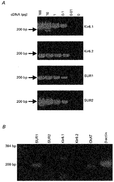
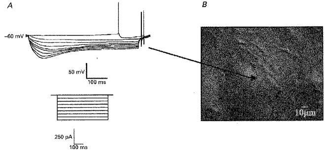
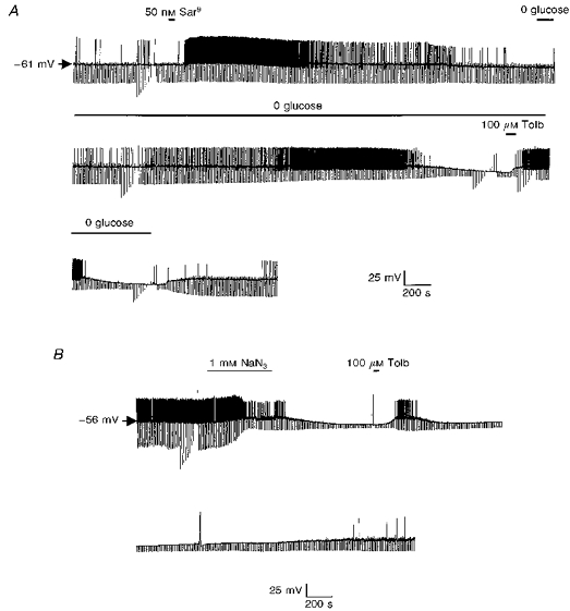
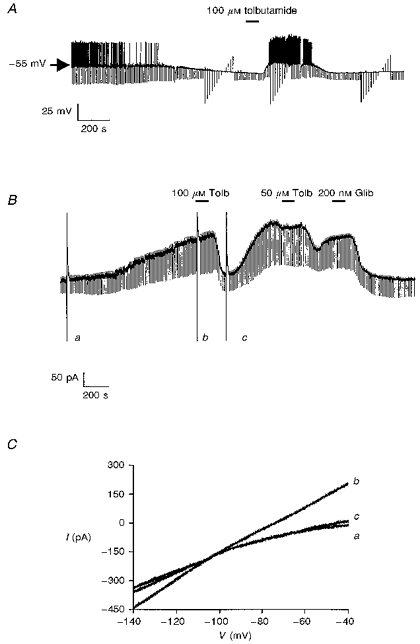
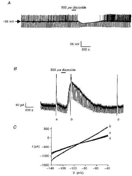
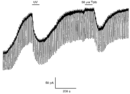
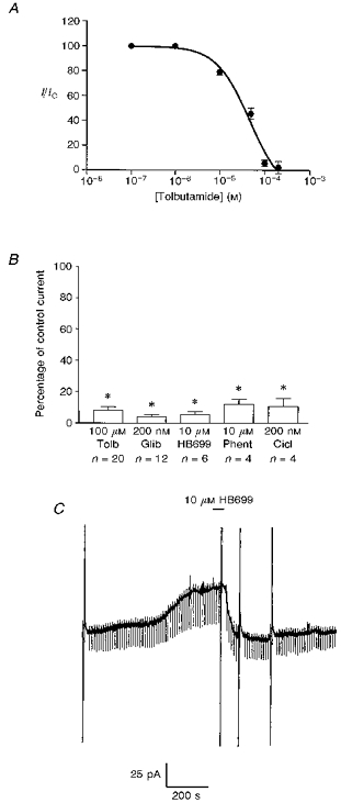
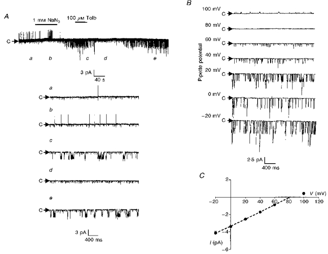
1. Whole-cell patch-clamp recordings were made from rat striatal cholinergic interneurones in slices of brain tissue in vitro. In the absence of ATP in the electrode solution, these neurones were found to gradually hyperpolarize through the induction of an outward current at -60 mV. This outward current and the resultant hyperpolarization were blocked by the sulphonylureas tolbutamide and glibenclamide and by the photorelease of caged ATP within neurones. 2. This ATP-sensitive outward current was not observed when 2 mM ATP was present in the electrode solution. Under these conditions, 500 microM diazoxide was found to induce an outward current that was blocked by tolbutamide. 3. Using permeabilized patch recordings, neurones were shown to hyperpolarize in response to glucose deprivation or metabolic poisoning with sodium azide (NaN3). The resultant hyperpolarization was blocked by tolbutamide. 4. In cell-attached recordings, metabolic inhibition with 1 mM NaN3 revealed the presence of a tolbutamide-sensitive channel exhibiting a unitary conductance of 44.1 pS. 5. Reverse transcription followed by the polymerase chain reaction using cytoplasm from single cholinergic interneurones demonstrated the expression of the ATP-sensitive potassium (KATP) channel subunits Kir6.1 and SUR1 but not Kir6.2 or SUR2. 6. It is concluded that cholinergic interneurones within the rat striatum exhibit a KATP channel current and that this channel is formed from Kir6.1 and SUR1 subunits.


Figures

Figure 8
Figure 8. Molecular identity of the KATP channel complex
A, 2.5%agarose gel showing the sensitivity of the Kir6.1, Kir6.2, SUR1 and SUR2 primer pairs. B, using the cytoplasmic contents harvested from a single cholinergic neurone, the expression of ChAT, Kir6.1, SUR1 and β-actin was detected.
Figure 1
Figure 1. Characterization of rat striatal cholinergic interneurones
A, voltage response of a cholinergic interneurone to hyperpolarizing current injection. Inset depicts current protocol. B, picture of a section of the striatum as viewed under infrared optics illustrating the appearance of a cholinergic interneurone (denoted by arrow).
Figure 6
Figure 6. The effect of aglycaemia and metabolic inhibition of the electrical activity of cholinergic interneurones
A, current-clamp recording using an amphotericin B-permeabilized patch. Application of the NK1 agonist Sar9 produced marked depolarization with associated increase in firing rate. Removal of glucose causes cellular hyperpolarization in a tolbutamide-sensitive manner. B, in this permeabilized patch recording metabolic inhibition with sodium azide (NaN3) hyperpolarized the cell and this was reversed by tolbutamide.
Figure 2
Figure 2. The effect of dialysis with an ATP-free electrode solution
A, continuous whole-cell current-clamp recording demonstrating the effect of tolbutamide on the hyperpolarization resulting from dialysis with an ATP-free electrode solution. B, the effect of tolbutamide (Tolb) and glibenclamide (Glib) on the time-dependent outward current at a holding potential of −60 mV. The large deflections are the current responses to voltage ramps from −140 to −40 mV as depicted in C. C, expanded traces of the current responses to the voltage ramps shown in B. The letters beside these currents correspond to those shown in B.
Figure 3
Figure 3. The effect of diazoxide on cholinergic interneurones
A, in the presence of 2 mM ATP in the electrode, diazoxide induces a membrane hyperpolarization with associated decrease in input resistance. B, in a neurone voltage clamped at −60 mV, diazoxide induces an outward current. The large deflections are the current responses to voltage ramps from −140 to −40 mV as depicted in C. C, expanded traces of the current responses to the voltage ramps shown in B. The letters beside these currents correspond to those shown in B.
Figure 4
Figure 4
UV photolysis of caged ATP inhibits the outward sulphonylurea-sensitive current in a cholinergic interneurone voltage clamped at −60 mV.
Figure 5
Figure 5. Pharmacology of the sulphonylurea-sensitive current
A, concentration-response curve for tolbutamide. All points are means of three to six separate experiments. Vertical lines indicate s.e.m.; where no lines are apparent, the error was within the size of the symbol. The associated line is the line of best fit to eqn (1). B, mean data illustrating the pharmacology of the sulphonylurea-sensitive current at −60 mV. The effect of each agent was compared with the control response obtained in the same neurone. HB699 (meglitinide); Phent, phentolamine; Cicl, ciclazindol. Error bars indicate s.e.m. and n shows the number of cells tested for each condition * Significantly less than the control response (100 %), P < 0.05. C, the effect of HB699 in a cell voltage clamped at −60 mV. The large vertical deflections are current responses to voltage ramps.
Figure 7
Figure 7. Single channel characteristics of the tolbutamide-sensitive channel activated by metabolic inhibition with NaN3
A, cell-attached recording made at a pipette potential of 0 mV. Bath application of NaN3 induced the activity of several 3.2 pA channels which were inhibited by tolbutamide. The letters refer to the panels shown on the expanded timescale in the lower part of the figure. Note the presence of a smaller channel that was active throughout the recording and was unaffected by tolbutamide. B, cell-attached recording of the NaN3-activated channel at different pipette potentials. C, current-voltage relationship revealed a unitary conductance of 41.4 ± 0.8 pS in the linear part of the plot (denoted by the dashed line). Each point is the mean of between three and five observations.

Similar articles

Cited by

References

    1. Aosaki T, Kawaguchi Y. Actions of substance P on rat neostriatal neurons in vitro. Journal of Neuroscience. 1996;16:5141–5153. - PMC - PubMed
    1. Ashcroft SJH, Ashcroft FM. Properties and functions of ATP-sensitive K+ channels. Cellular Signalling. 1990;2:197–214. 10.1016/0898-6568(90)90048-F. - DOI - PubMed
    1. Calabresi P, Ascone CM, Centonze D, Pisani D, Sancesario G, D'Angelo V, Bernardi G. Opposite membrane potential changes induced by glucose deprivation in striatal spiny neurons and in large aspiny interneurons. Journal of Neuroscience. 1997;17:1940–1949. - PMC - PubMed
    1. Chesselet M-F, Gonzales C, Lin C-S, Polsky K, Jin B-K. Ischemic damage in the striatum of adult gerbils: Relative sparing of somatostatinergic and cholinergic interneurons contrasts with loss of efferent neurons. Experimental Neurology. 1990;110:209–218. - PubMed
    1. Di Chiara G, Morelli M, Consolo S. Modulatory functions of neurotransmitters in the striatum: ACh/dopamine/ NMDA interactions. Trends in Neurosciences. 1994;17:228–233. 10.1016/0166-2236(94)90005-1. - DOI - PubMed

MeSH terms

LinkOut - more resources