. 1998 Sep 1;511 ( Pt 2)(Pt 2):519-31.
doi: 10.1111/j.1469-7793.1998.519bh.x.

Force-velocity and power-load curves in rat skinned cardiac myocytes

Affiliations

Force-velocity and power-load curves in rat skinned cardiac myocytes

K S McDonald et al. J Physiol. .

Abstract

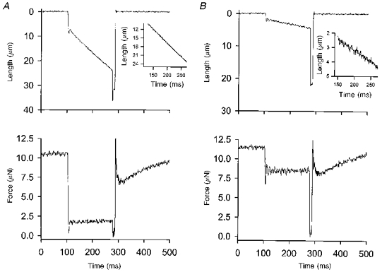
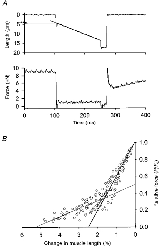
1. This study utilized a skinned myocyte preparation with low end compliance to examine force-velocity and power-load curves at 12 C in myocytes from rat hearts. 2. In maximally activated myocyte preparations, shortening velocities appeared to remain constant during load clamps in which shortening took place over a sarcomere length range of approximately 2.30-2.00 micro m. These results suggest that previously reported curvilinear length traces during load clamps of multicellular preparations were due in part to extracellular viscoelastic structures that give rise to restoring forces during myocardial shortening. 3. During submaximal Ca2+ activations, the velocity of shortening at low loads slowed and the time course of shortening became curvilinear, i.e. velocity progressively slowed as shortening continued. This result implies that cross-bridge cycling kinetics are slower at low levels of activation and that an internal load arises during shortening of submaximally activated myocytes, perhaps due to slowly detaching cross-bridges. 4. Reduced levels of activator Ca2+ also reduced maximal power output and increased the relative load at which power output was optimal. For a given absolute load, the shift has the effect of maintaining power output near the optimum level despite reductions in cross-bridge number and force generating capability at lower levels of Ca2+.


Figures

Figure 1
Figure 1. Photomicrographs of a single rat skinned cardiac myocyte preparation
A, relaxed myocyte (pCa 9.0); B, during maximal activation (pCa 4.5). As illustrated in the drawing (C, modified from Fig. 3, Metzger, Greaser & Moss, 1989), the myocyte preparation was mounted between a force transducer and a position motor. The ends of the myocyte were secured by overlaying a 0.5 mm piece of 4–0 suture, which were tied into troughs using loops of 10–0 suture. The length of myocyte between the two troughs in this preparation is 160 μm. D shows force traces during maximal activation of this myocyte preparation following a rapid change (Δ) in myocyte length. The maximal force generated by this preparation was 9.5 μN.
Figure 3
Figure 3. Length traces during isotonic shortening in a maximally activated myocyte preparation
A shows length (top) and force (bottom) traces during an isotonic clamp to 0.22 of peak force (Po); the inset shows an enlargement of the length trace during isotonic shortening. The straight line fit is superimposed on the length trace. B shows a load clamp to 0.75 Po in the same myocyte. In A and B, the myocyte preparation was rapidly slackened after the period of isotonic shortening and after 20 ms the preparation was rapidly relengthened to its original length.
Figure 2
Figure 2. Estimation of myocyte preparation end compliance
A, representative myocyte length (ML) trace and force clamp during isotonic shortening. The length change (indicated by the asterisk) associated with reducing force to the level shown was estimated by extrapolating the fitted straight line to the onset of the force clamp. The oscillation in the length trace prior to isotonic shortening is due to initial overshoot in the force clamp induced by the feedback circuit. B, plot of instantaneous length change versus relative load at the end of the length step. The relationship exhibits two phases, a linear phase for relative forces (P/Po) between 1.0 and 0.5 Po and a curvilinear phase for forces < 0.5 Po. The linear phase was used to estimate series elasticity of the myocyte preparations. Extrapolation of the linear phase approximates a series elasticity of 2.5% myocyte preparation length.
Figure 4
Figure 4. Myocyte length (ML), sarcomere length (SL), and tension during a load clamp
A, both ML and SL exhibited linear shortening time courses during the load clamp. The regression coefficients for ML and SL were 0.996 and 0.985, respectively. B shows force-velocity points for both whole preparation shortening (○) and sarcomere shortening (•) during the same load clamps, indicating that changes in myocyte preparation length during isotonic shortening closely reflect shortening at the level of the sarcomere.
Figure 5
Figure 5. Cumulative force-velocity (•) and power output (○) relationships
Data were obtained from 13 skinned rat cardiac myocyte preparations during maximal activation. Data points in both force-velocity and power-load curves are means ±s.d. Mean force-velocity data were fitted using the normalized form of the Hill equation: where Vmax and Po are maximum velocity of shortening and isometric tension, respectively, and a is a force constant.
Figure 6
Figure 6. Effects of Ca2+ on shortening during isotonic load clamps in a rat skinned cardiac myocyte preparation
The myocyte preparation shortened at a constant velocity during the entire load clamp during maximal activation (i.e. pCa 4.5). When [Ca2+] was reduced to yield ≈50%Po (i.e. pCa 5.8), the length trace was curvilinear during isotonic shortening. The length trace was fitted using a single exponential equation. The dashed line illustrates the deviation from linearity of the length trace. The dashed line was best-fitted by eye for ≈50 ms of the length trace near the beginning of the load clamp.
Figure 7
Figure 7. Plots of force-velocity and power-load data obtained during maximal (pCa 4.5) and submaximal activations (P/Po = 0.49 ± 0.04)
Plotted values are pooled results from 7 myocyte preparations. In A and B, the load at each step was expressed as a fraction of isometric tension for each myocyte preparation. In C, the load was normalized to force during maximal activation (i.e. pCa 4.5). For submaximal activations, length traces during load clamps were well fitted by a single exponential equation. Initial velocities of shortening at time 0 ms (i.e. immediately after the onset of the load clamp) are plotted in A. Shortening velocities at 100 ms following initiation of the load clamp are also plotted in A and these velocities were used to calculate power outputs, which are plotted in B and C.
Figure 8
Figure 8. Schematic representation of the effect of contractility on force-velocity curves and its implications for myocardial performance
A, force-velocity curves are shown for a myocyte during maximal and submaximal Ca2+ activation. An elevation in [Ca2+] doubles the maximum force-generating capacity (i.e. force increases from 0.5 to 1.0 P4.5) and increases the curvature of the force-velocity relationship. Since force increased 2-fold, an after-load that was 40% of isometric force at low [Ca2+] force becomes only 20% of isometric force produced at high [Ca2+]. B shows power-load curves normalized to low [Ca2+] force and high [Ca2+] force. The force optimal for power output is 0.40 of low [Ca2+] isometric force and 0.20 of high [Ca2+] isometric force as a result of alterations in the shape of the force-velocity curves. C also shows power-load curves at low and high [Ca2+], but in this case normalized only to high [Ca2+] force. This illustrates that power output is optimal at the same absolute load during both low and high [Ca2+] activations. Thus, when myocardium is working against the same after-load, changes in force-velocity properties in response to varying [Ca2+] have the effect of maintaining power output near optimal levels despite altered contractility.

Similar articles

Cited by

References

    1. Brady AJ. Mechanical properties of isolated cardiac myocytes. Biological Reviews. 1991;71:413–428. - PubMed
    1. Brenner B. Calcium activation and maximum unloaded shortening velocity. Investigations on glycerinated skeletal and heart muscle preparations. Basic Research in Cardiology. 1980;75:40–46. - PubMed
    1. Brenner B. The necessity of using two parameters to describe isotonic shortening velocity of muscle tissues: The effect of various interventions upon initial shortening velocity (vi) and curvature (b) Basic Research in Cardiology. 1986;81:54–69. - PubMed
    1. Brutsaert DL, Claes VA, Goethals MA. Effect of calcium on force-velocity relations of heart muscle of the cat. Circulation Research. 1973;32:385–392. - PubMed
    1. Chiu YC, Ballou EW, Ford LE. Internal viscoelastic loading in cat papillary muscle. Biophysical Journal. 1982;40:109–120. - PMC - PubMed

Publication types

LinkOut - more resources