. 1998 Sep 1;511 ( Pt 2)(Pt 2):629-43.
doi: 10.1111/j.1469-7793.1998.629bh.x.

Effect of depletion of glycosaminoglycans and non-collagenous proteins on interstitial hydraulic permeability in rabbit synovium

Affiliations

Effect of depletion of glycosaminoglycans and non-collagenous proteins on interstitial hydraulic permeability in rabbit synovium

D Scott et al. J Physiol. .

Abstract

1. The hydraulic resistance of synovial interstitium helps to retain a lubricating fluid within the joint cavity. The contributions of sulphated glycosaminoglycans to resistance were assessed by selective depletion by chondroitinase ABC, keratanase and heparinases I, II and III in vivo. Also, since glycosaminoglycans do not account fully for the resistance, the contribution of non-collagenous, structural proteins in interstitium was assessed by treatment with chymopapain, a collagen-sparing protease. 2. Ringer solution containing enzyme was injected into the synovial cavity of the knee in anaesthetized rabbits. After >= 30 min the intra-articular pressure was raised and the relation between pressure (Pj) and trans-synovial outflow (Qs) determined. The slope dQs/dPj at low pressures, i.e. below yield pressure, represents the hydraulic conductance of the lining, i.e. 1/resistance. The contralateral joint received Ringer solution without enzyme as a control. Action of enzymes on the tissue was confirmed by histochemical and immunohistochemical studies. 3. Treatment with chondroitinase ABC (5 joints) increased the hydraulic conductance of the lining by 2.3 times (control, 1.34 +/- 0.22 microliter l min-1 cmH2O-1; post-enzyme, 3.11 +/- 0.45 microliter l min-1 cmH2O-1). This was significantly less than the effects of leech, Streptomyces and testicular hyaluronidases, which caused an average 4.7 times increase (P < 0.001, ANOVA). Analogous findings were made above yield pressure. 4. Treatment with keratanase (3 joints) or heparinases I, II and III (3 joints) caused no significant increase in trans-synovial flows or conductance, even though the concentration of heparan sulphate in synovium is higher than that of chondroitin sulphates or hyaluronan. 5. Treatment with chymopapain (7 joints) caused the greatest increases in trans-synovial flow, which exceeded control flow by an order of magnitude in one case. After 0.1 U chymopapain the average conductance was 6.6 times the control conductance below yield pressure. Immunohistochemical studies confirmed that chymopapain treatment removed the synovial proteoglycans. 6. It is concluded that, despite their similar resistivities in vitro, the different glycosaminoglycans do not contribute equally, weight for weight, to interstitial resistance in vivo. Hyaluronan is the dominant glycosaminoglycan governing synovial interstitial resistance. In addition, non-collagenous structural proteins contribute significantly to interstitial resistance.


Figures

Figure 1
Figure 1. Photomicrographs of synovium (S), subsynovium (SS) and underlying muscle (M) from the suprapatellar bursa of the knee joint cavity (JC), showing effects of treatment by glycosaminoglycanidases
A, control synovium showing normal distribution of hyaluronan (HABR preparation). B, synovium treated with chondroitinase ABC in vivo. Only small amounts of hyaluronan remain (HABR preparation). The hydraulic conductance of this tissue, measured prior to sampling, was raised. C, control synovium showing normal distribution of keratan sulphate (5-D-4 antibody). D, synovium treated with keratanase in vivo, showing absence of keratan sulphate (5-D-4 antibody). Synovial hydraulic conductance was not significantly changed. All photomicrographs are at the same magnification; bar in A, 10 μm.
Figure 6
Figure 6. Effect of chymopapain on synovial structure and immunohistochemistry
A, the normal, control synovium (S) is a thin layer of cells and interstitium with small blood vessels (arrows). The underlying subsynovium (SS) is a loose connective tissue with few cells. It lies on muscle (M). (Haematoxylin and Eosin staining.) B, synovium and subsynovium after treatment with 0.1 U chymopapain in vivo are oedematous and thickened. The cells are more widely spaced and the blood vessels (arrows) are enlarged. The hydraulic conductance of this tissue, measured prior to excision, was greatly increased. (Haematoxylin and Eosin staining.) C, synovium after treatment with 10 U chymopapain in vivo is disrupted. The cells that remain on the surface appear to be embedded in a hyaline layer (arrows). The subsynovium is filled with red cells. The blood vessels (BV) are very swollen. (Haematoxylin and Eosin staining.) D, after treatment with 0.1 U chymopapain in vivo much of the hyaluronan is removed from the synovium (HABR preparation). Compare with Fig. 1A. E, control synovium, showing the distribution of chondroitin 6-sulphate oligosaccharides (3-B-3 antibody). F, after treatment with 0.1 U chymopapain in vivo, little if any chondroitin 6-sulphate remains (3-B-3 antibody). All photomicrographs are at the same magnification; bar in A, 10 μm.
Figure 2
Figure 2. Effect of chondroitinase ABC on the rate of absorption of Ringer solution from the cavity of the rabbit knee
A, results from contralateral knees of the same rabbit. One joint was pre-treated for 30 min with 10 U chondroitinase ABC in Ringer solution (•) and the other with plain Ringer solution (○), prior to determining the pressure-flow relation. Dashed lines show regressions through the results in the low and high pressure ranges (below and above yield pressure). B, comparison of interpolated flows at the same intra-articular pressure, with (formula image) or without chondroitinase ABC pre-treatment (□) in pairs of knees from five rabbits (means ±s.e.m.). Statistical significance is indicated (*P≤ 0.05, **P≤ 0.01, ***P < = 0.001; Student's paired t test).
Figure 3
Figure 3. Effect of keratanase treatment on rate the of absorption of Ringer solution from the cavity of the rabbit knee
A, results from contralateral knees of the same rabbit. One was pre-treated for 30 min with keratanase in Ringer solution (•) and the other with plain Ringer solution (○) prior to determination of the pressure-flow relation. Note expanded scale of the y-axis compared with Fig. 2. Dashed lines are regressions through the results below and above yield pressure. B, comparison of interpolated flows at the same intra-articular pressure with (formula image) or without keratanase pre-treatment (□) in pairs of knees from three rabbits (means ±s.e.m.), plotted on the same y-axis scale as Fig. 2 for comparison. None of the differences between control and keratanase-treated joints are statistically significant.
Figure 4
Figure 4. Effect of chymopapain treatment on the rate of absorption of Ringer solution from the synovial cavity of rabbit knees
The scale on the y-axis is contracted relative to Figs 2 and 3 in order to accommodate the very large flows. A, synovium injected with 0.1 U chymopapain, 90 min before measuring the pressure-flow relation. Dashed lines are regressions through the results below and above yield pressure. B, comparison of interpolated flows at same intra-articular pressures (means ± s.e.m) after chymopapain pre-treatment (formula image) and in control joints (□; Student's t test, *P≤ 0.05, **P < = 0.01).
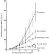
Figure 5
Figure 5. Comparison of the effects of various glycosaminoglycanidases and a protease, chymopapain, on the pressure-flow relation of the synovial lining
The keratanase results (Fig. 3) are not shown because they almost exactly overlie the heparinase results. The hyaluronidase results are the means of the pooled results for testicular, Streptomyces and leech hyaluronidases from Coleman et al. (; n = 14). The control values are the means of the pooled control flows from the chondroitinase ABC, keratanase, heparinase and chymopapain studies (n = 16). Results shown are means ±s.e.m.
Figure 7
Figure 7. Comparison of observed effects of glycosaminoglycanidases and protease on synovial hydraulic conductance (data points, means ±s.e.m.) with predictions of a mathematical model (see text)
The vertical axis is hydraulic conductance below yield pressure after enzyme treatment, i.e. after partial depletion of biopolymer, relative to control conductance. The dot-and-dashed horizontal line marks the average, 6.6-fold increase in conductance caused by treatment with 0.1 U chymopapain, to 10.8 μl min−1 cmH2O−1 (below yield pressure). This corresponds to a residual extrafibrillar biopolymer concentration of 6 mg ml−1 (vertical line).

Similar articles

Cited by

References

    1. Bert JL, Reed RK. Hyaluronan and the flow conductivity of rat dermis. In: Reed RK, Rubin K, editors. Connective Tissue Biology, Integration and Reductionism. London: Portland Press; 1998. pp. 41–48.
    1. Bradford DS, Oegema TR, Cooper KM, Wakano K, Chao EY. Chymopapain, chemonucleolysis and nucleus pulposus regeneration. A biochemical and biomechanical study. Spine. 1984;9:135–147. - PubMed
    1. Buchmann MD, Gluzband YA, Grodzinsky AJ, Hunzicker EB. Mechanical compression modulates matrix biosynthesis in chondrocyte/agarose culture. Journal of Cell Science. 1995;108:1497–1508. - PubMed
    1. Caterson B, Christner JE, Baker JR, Couchman D. Production and characterization of monoclonal antibodies directed against connective tissue proteoglycans. Federation Proceedings. 1985;44:386–393. - PubMed
    1. Coleman PJ, Kavanagh E, Mason RM, Levick JR, Ashhurst DE. The proteoglycans and glycosaminoglycan chains of rabbit synovium. Histochemical Journal. 1998a in the Press. - PubMed

Publication types

LinkOut - more resources