. 1998 Aug 18;95(17):9755-60.
doi: 10.1073/pnas.95.17.9755.

Biochemical basis of SOS-induced mutagenesis in Escherichia coli: reconstitution of in vitro lesion bypass dependent on the UmuD'2C mutagenic complex and RecA protein

Affiliations

Biochemical basis of SOS-induced mutagenesis in Escherichia coli: reconstitution of in vitro lesion bypass dependent on the UmuD'2C mutagenic complex and RecA protein

M Tang et al. Proc Natl Acad Sci U S A. .

Abstract

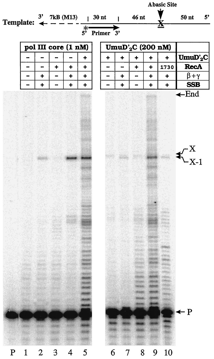
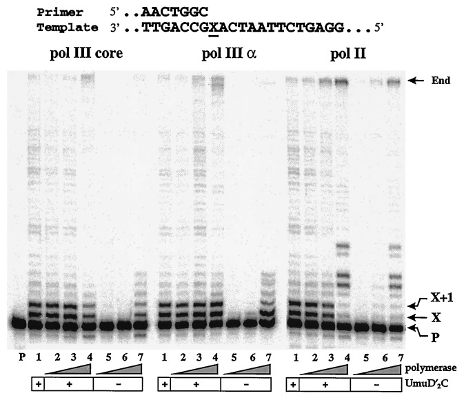
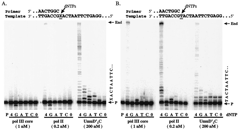
Damage-induced SOS mutagenesis requiring the UmuD'C proteins occurs as part of the cells' global response to DNA damage. In vitro studies on the biochemical basis of SOS mutagenesis have been hampered by difficulties in obtaining biologically active UmuC protein, which, when overproduced, is insoluble in aqueous solution. We have circumvented this problem by purifying the UmuD'2C complex in soluble form and have used it to reconstitute an SOS lesion bypass system in vitro. Stimulated bypass of a site-directed model abasic lesion occurs in the presence of UmuD'2C, activated RecA protein (RecA*), beta-sliding clamp, gamma-clamp loading complex, single-stranded binding protein (SSB), and either DNA polymerases III or II. Synthesis in the presence of UmuD'2C is nonprocessive on damaged and undamaged DNA. No lesion bypass is observed when wild-type RecA is replaced with RecA1730, a mutant that is specifically defective for Umu-dependent mutagenesis. Perhaps the most noteworthy property of UmuD'2C resides in its ability to stimulate both nucleotide misincorporation and mismatch extension at aberrant and normal template sites. These observations provide a biochemical basis for the role of the Umu complex in SOS-targeted and SOS-untargeted mutagenesis.


Figures

Figure 1
Figure 1
UmuD′2C-stimulated abasic site bypass. Standard polymerization reactions, using a running-start protocol, were carried out in the presence or absence of exogenous pol III core by using combinations of UmuD′2C, RecA, β,γ-complex, and SSB. Four dNTPs (100 μM) and ATP (1 mM) were present in all reactions. A 32P-labeled primer was annealed to a DNA template containing an abasic lesion, X (top of figure), and the replication products were separated in 10% denaturing polyacrylamide gels and visualized by phosphorimaging. Locations of the unextended primer band, abasic site (X), upstream site adjacent to the lesion (X − 1), and the end of template are indicated on the right. Lane P contains the primer in the absence of proteins. Additions to the replication reaction mixtures are shown in the box at the top of the gel; β + γ, represents the β-clamp processivity subunit of the pol III holoenzyme complex and the five protein γ-clamp loading complex consisting of the subunits γ, δ, δ′, χ, ψ.
Figure 2
Figure 2
Effect of pol III and pol II on translesion replication. Standard polymerization reactions, using a standing-start protocol, were carried out in either the presence or absence of UmuD′2C by using different concentrations of pol III core (0, 0.5, 2, 20 nM), pol III α-subunit (0, 0.5, 2, 20 nM), and pol II (0, 0.2, 1, 10 nM). All reactions contain RecA, β,γ-complex, SSB, four dNTPs (100 μM), and ATP (1 mM). Lane P contains the 32P-labeled primer in the absence of proteins. Locations of the unextended primer band, abasic site (X), downstream site adjacent to the lesion (X + 1), and end of template are indicated on the right. The DNA used in the standing-start protocol is shown at the top.
Figure 3
Figure 3
Effect of UmuD′2C on nucleotide misincorporation and mismatch extension on lesion-containing and normal DNA templates. Standard standing-start polymerization reactions were carried out by using pol III core, pol II, or UmuD′2C. (A) Reactions carried out by using a DNA template containing an abasic lesion, X. (B) Reactions carried out by using a natural DNA template in which X is replaced by T. The lanes labeled as G, A, T, and C denote reactions carried out with a single dNTP substrate, dGTP, dATP, dTTP, and dCTP, respectively. The lanes labeled as 4 and 0 denote reactions carried out in the presence and absence of four dNTPs, respectively. Lane P contains the 32P-labeled primer in the absence of proteins. The abasic lesion containing and natural DNA templates are shown above each gel. A portion of each template sequence is shown on the right. UmuD′2C measurements made in the presence of pol III (1 nM) and pol II (0.2 nM), resulted in banding patterns identical to those shown in the UmuD′2C panel.

Similar articles

Cited by

References

    1. Witkin E M. Bacteriol Rev. 1976;40:869–907. - PMC - PubMed
    1. Walker G C. Microbiol Rev. 1984;48:60–93. - PMC - PubMed
    1. Friedberg E C, Walker G C, Siede W. DNA Repair and Mutagenesis. Washington, DC: Am. Soc. Microbiol.; 1995.
    1. Echols H, Goodman M F. Annu Rev Biochem. 1991;60:477–511. - PubMed
    1. Koffel-Schwartz N, Coin F, Veaute X, Fuchs R P P. Proc Natl Acad Sci USA. 1996;93:7805–7810. - PMC - PubMed

Publication types

MeSH terms

LinkOut - more resources