. 1998 Aug 18;95(17):9779-84.
doi: 10.1073/pnas.95.17.9779.

PDZ-domain-mediated interaction of the Eph-related receptor tyrosine kinase EphB3 and the ras-binding protein AF6 depends on the kinase activity of the receptor

Affiliations

PDZ-domain-mediated interaction of the Eph-related receptor tyrosine kinase EphB3 and the ras-binding protein AF6 depends on the kinase activity of the receptor

B Hock et al. Proc Natl Acad Sci U S A. .

Abstract

Eph-related receptor tyrosine kinases (RTKs) have been implicated in intercellular communication during embryonic development. To elucidate their signal transduction pathways, we applied the yeast two-hybrid system. We could demonstrate that the carboxyl termini of the Eph-related RTKs EphA7, EphB2, EphB3, EphB5, and EphB6 interact with the PDZ domain of the ras-binding protein AF6. A mutational analysis revealed that six C-terminal residues of the receptors are involved in binding to the PDZ domain of AF6 in a sequence-specific fashion. Moreover, this PDZ domain also interacts with C-terminal sequences derived from other transmembrane receptors such as neurexins and the Notch ligand Jagged. In contrast to the association of EphB3 to the PDZ domain of AF6, the interaction with full-length AF6 clearly depends on the kinase activity of EphB3, suggesting a regulated mechanism for the PDZ-domain-mediated interaction. These data gave rise to the idea that the binding of AF6 to EphB3 occurs in a cooperative fashion because of synergistic effects involving different epitopes of both proteins. Moreover, in NIH 3T3 and NG108 cells endogenous AF6 is phosphorylated specifically by EphB3 and EphB2 in a ligand-dependent fashion. Our observations add the PDZ domain to the group of conserved protein modules such as Src-homology-2 (SH2) and phosphotyrosine-binding (PTB) domains that regulate signal transduction through their ability to mediate the interaction with RTKs.


Figures

Figure 1
Figure 1
Interaction of the PDZ domain of AF6 with the C termini of EphB2 and EphB3. (A) AF6-domain topology: ras-binding domain (RBD), homology regions to kinesin-like proteins (U104) and to class V myosins (DIL), PDZ domain (PDZ). cDNAs identified in a two-hybrid screening are shown relative to the enlarged PDZ domain. (B) Intracellular domains of EphB2 and EphB3 containing deletions or point mutations were tested in two-hybrid assays for their ability to bind to the PDZ domain of AF6. Interactions were detected by induction of HIS3 expression on medium lacking histidine. KD refers to the wild-type receptor, KD- to the kinase-deficient receptor. Sequences of C-terminal peptides are indicated. The C-terminal elongation of EphB3 by 15 aa is shown in black.
Figure 2
Figure 2
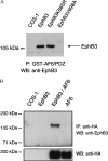
Association of EphB3 and the PDZ domain of AF-6 in vitro and in mammalian cells. (A) Wild-type or mutant EphB3 was expressed in COS-1 cells as indicated. Proteins that could be precipitated with the GST-AF6/PDZ fusion (P) were treated on a Western blot (WB) with EphB3 antibodies. (B) Lysates from untransfected, EphB3-transfected, EphB3- and HA-AF6-cotransfected, or HA-AF6-transfected COS-1 cells were immunoprecipitated (IP) with HA antibodies. Immunoprecipitates were probed on a Western blot (WB) with EphB3 antiserum, stripped, and reprobed with monoclonal HA antibodies.
Figure 3
Figure 3
Phosphorylation of full-length AF6 depends on the catalytic activity of the receptor. (A) COS-1 cells were cotransfected with various mutants of EphB3 and HA-AF6. Cell lysates were probed on a Western blot with phosphotyrosine antibodies. AF6 is recognized as multiple bands presumably due to degradation of the overexpressed protein. The EphB3 and HA-AF6 content of the lysates was compared on separate blots using HA (not shown) and EphB3 antisera (Lower). (B) NIH 3T3, NG108, or NG108 cells stably expressing EphB2 (NG108/NUK) were challenged with 2 μg/ml clustered ephrine-B1 Fc (+) or human Fc antibodies (Sigma) for 30 min (−). Subsequently, cells were scraped off, washed twice in PBS, and lysed in RIPA buffer. Cell lysates were immunoprecipitated (IP) with a polyclonal AF6 antiserum and probed on a Western blot (WB) using monoclonal phosphotyrosine antibodies (PY99, Santa Cruz Biotechnology), stripped, and reprobed with AF6 antibodies.
Figure 4
Figure 4
EphB3 binding to full-length AF6 depends on the catalytic activity of the receptor. COS-1 cells were cotransfected with various mutants of EphB3 and HA-AF6. Cell lysates were immunoprecipitated (IP) with HA antibodies and probed on a Western blot (WB) using EphB3 antisera, stripped, and reprobed with monoclonal HA antibodies.
Figure 5
Figure 5
Northern blot analysis of different neuronal (A) and nonneuronal (B) human tissues. Blots were probed with a 291-bp fragment of the AF6 cDNA encompassing the PDZ domain under high-stringency conditions. After exposure blots were stripped and reprobed with an EphB3-specific cDNA probe under the same conditions.

Similar articles

Cited by

References

    1. van der Geer P, Hunter T, Lindberg R A. Annu Rev Cell Biol. 1994;10:251–337. - PubMed
    1. Pawson T. Nature (London) 1995;373:573–580. - PubMed
    1. Böhme B, Holtrich U, Wolf G, Luzius H, Grzeschik K H, Strebhardt K, Rübsamen-Waigmann H. Oncogene. 1993;8:2857–2862. - PubMed
    1. Eph Nomenclature Committee. Cell. 1997;90:403–404. - PubMed
    1. Böhme B, VandenBos T, Cerretti D P, Park L S, Holtrich U, Rübsamen-Waigmann H, Strebhardt K. J Biol Chem. 1996;271:24747–24752. - PubMed

Publication types

MeSH terms

LinkOut - more resources