. 1998 Aug 18;95(17):9813-8.
doi: 10.1073/pnas.95.17.9813.

New enzyme lineages by subdomain shuffling

Affiliations

New enzyme lineages by subdomain shuffling

K P Hopfner et al. Proc Natl Acad Sci U S A. .

Abstract

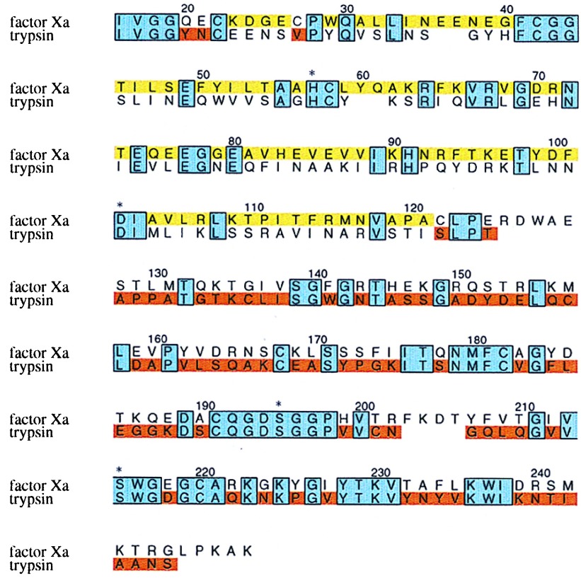
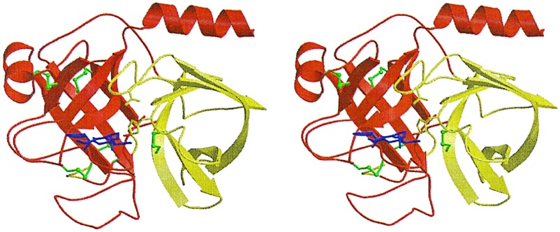
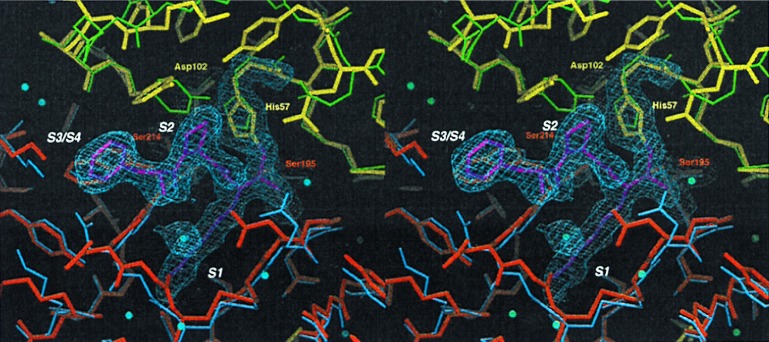
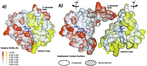
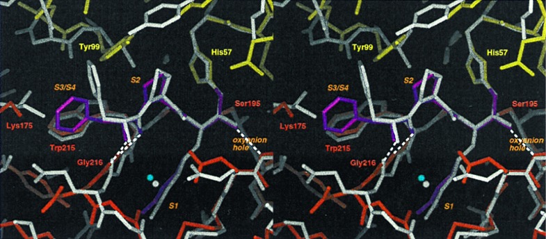
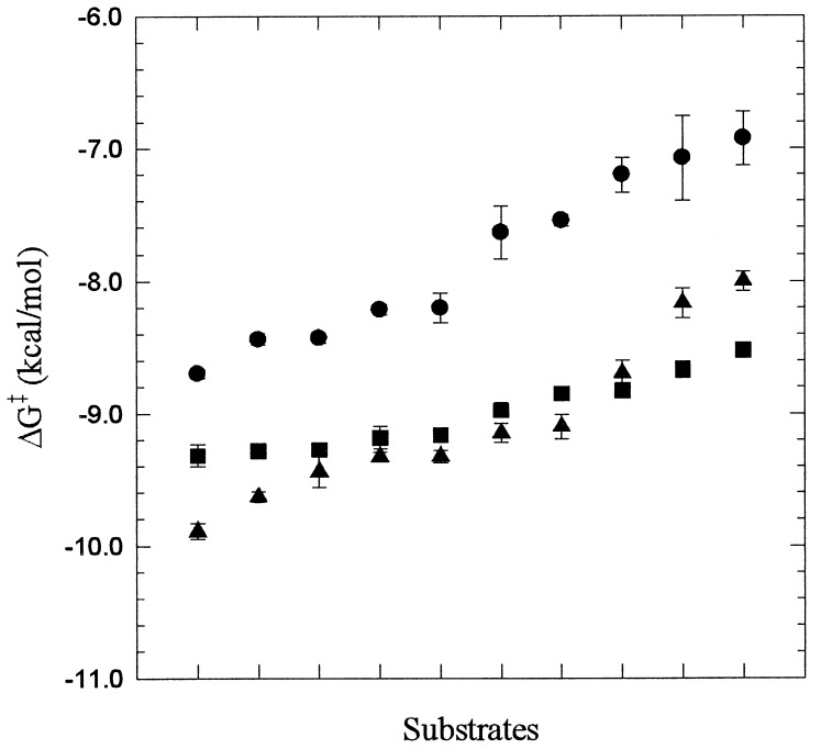
Protein functions have evolved in part via domain recombination events. Such events, for example, recombine structurally independent functional domains and shuffle targeting, regulatory, and/or catalytic functions. Domain recombination, however, can generate new functions, as implied by the observation of catalytic sites at interfaces of distinct folding domains. If useful to an evolving organism, such initially rudimentary functions would likely acquire greater efficiency and diversity, whereas the initially distinct folding domains would likely develop into single functional domains. This represents the probable evolution of the S1 serine protease family, whose two homologous beta-barrel subdomains assemble to form the binding sites and the catalytic machinery. Among S1 family members, the contact interface and catalytic residues are highly conserved whereas surrounding surfaces are highly variable. This observation suggests a new strategy to engineer viable proteins with novel properties, by swapping folding subdomains chosen from among protein family members. Such hybrid proteins would retain properties conserved throughout the family, including folding stability as single domain proteins, while providing new surfaces amenable to directed evolution or engineering of specific new properties. We show here that recombining the N-terminal subdomain from coagulation factor X with the C-terminal subdomain from trypsin creates a potent enzyme (fXYa) with novel properties, in particular a broad substrate specificity. As shown by the 2.15-A crystal structure, plasticity at the hydrophobic subdomain interface maintains activity, while surface loops are displaced compared with the parent subdomains. fXYa thus represents a new serine proteinase lineage with hybrid fX, trypsin, and novel properties.


Figures

Figure 1
Figure 1
Primary structure of the fXa/trypsin hybrid. The primary structure of rfXYa is shown in an amino acid sequence alignment (single letter code) of the catalytic domain of human coagulation factor Xa and human trypsin 1. Conserved residues (38% sequence identity) are boxed and shaded blue. Segments taken from fXa are colored yellow; those from trypsin are colored red. Three residues at the N terminus (Y20,E21 and V27) were taken from trypsin (red) to account for a different disulfide bridge in fXa (C22-C27) and trypsin (C22-C157). The trypsin disulfide bridge C22-C157 was used. Both subdomains contribute to the catalytic tetrad (denoted by asterisk).
Figure 2
Figure 2
Crystal structure of the fXa/trypsin hybrid. Ribbon plot of the crystal structure of fXYa. The N-terminal subdomain is shown in red, and the C-terminal subdomain is shown in yellow. Both subdomains adopt a β-barrel fold and assemble asymmetrically to generate the fold typical of the chymotrypsin family. Disulfide bridges are depicted in green (the N-terminal bridge discussed in the text is located behind the C-terminal barrel). The D-Phe-Pro-Arg inhibitor is shown with magenta sticks. It is bound to the active site, which is formed at the subdomain interface. The catalytic triad residue side chains are displayed explicitly as sticks.
Figure 3
Figure 3
Comparison of the active site of fXYa to fXa and trypsin. Final model of fXYa showing the active site (thick sticks using the color code of Fig. 2) with representative 1.0 σ contoured 2Fc-Fo electron density for the catalytic tetrad residues His52, Asp102, Ser195, and Ser214 as well as for the PPACK inhibitor. The N-terminal subdomain of fXYa (red) is superimposed with the N-terminal subdomain of fXa (blue); the C-terminal subdomain (yellow) is superimposed with the C-terminal subdomain of trypsin (green). The structure shows a well conserved catalytic triad and specificity pocket. Some side chain adjustments in substrate binding sites (S1-S3) presumably originate from interaction with PPACK.
Figure 4
Figure 4
Plasticity of the domain interface. (A) Connolly surface representation of rfXYa using the orientation and colors of Fig. 2. The surface was calculated for each subdomain separately. The extent of displacement of rfXYa surface atoms relative to the parent molecules after Ca superposition of the corresponding subdomains is color coded (Inset). PPACK (magenta) is shown as stick model. (B) Same as A but with N- and C-terminal subdomains rotated −30° and +30° about the vertical axis and separated to display the subunit interface. A stick model of the inhibitor (magenta) is displayed with each subdomain to clarify the respective orientation and to mark the location of the active site. The black bounded area denotes the contact surface of both subdomains; the shaded areas indicate surfaces of residues that differ between fX and trypsin. Core elements of the interface are structurally conserved. Surface patches of the interface are often deformed, especially at positions where nonconserved residues from both subdomains contact each other, such as the C-terminal helix and the calcium loop. These deviations involve both main chain and side chain geometry.
Figure 5
Figure 5
Inhibitor binding. The active site of PPACK-rfXYa (colored sticks using the code of Fig. 2) superimposed with the active site of PPACK-thrombin (grey sticks; PDB ID code 1ppb). PPACK binds in a substrate-like binding mode: P1-arginine extends into the S1-pocket; the backbone forms a β-sheet with the enzyme backbone at positions 214–216; and P2-proline occupies the S2-pocket. Characteristic hydrogen bonds of the transition state–serine proteinase interaction are formed (dashed lines). The binding mode is identical to that observed in PPACK thrombin except for the orientation of the P3 sidechain: in thrombin, the phenyl moiety of P3 fits into the S3/S4 site whereas in rfXYa it is rotated toward the bulk solvent.
Figure 6
Figure 6
Plot of the transition state stabilization free energy ΔG = −RTln(kcat/Km/knon) (35) for hydrolysis of substrates listed in Table 1 by rfXa (•), rfXYa (■), and rtrypsin (▴). The rate of the noncatalyzed reaction knon is independent of the enzyme used and was set to 1 for the purpose of the comparison. The SEs are shown as bars. To indicate selectivity, the substrates are sorted according the increasing ΔG values for each of the enzymes separately.

Similar articles

Cited by

References

    1. Bryson J W, Betz S F, Lu H S, Suich D J, Zhou H X, O’Neil K T, DeGrado W F. Science. 1995;270:935–941. - PubMed
    1. Lesk A M, Rose D R. Proc Natl Acad Sci USA. 1981;78:4304–4308. - PMC - PubMed
    1. Brinkmann U. Mol Med Today. 1996;2:439–446. - PubMed
    1. Chang J Y, Stafford D W, Straight D L. Biochemistry. 1995;34:12227–12232. - PubMed
    1. Neurath H. Science. 1984;224:350–357. - PubMed

Publication types

MeSH terms

Associated data