. 1998 Aug 18;95(17):9837-42.
doi: 10.1073/pnas.95.17.9837.

Primary activation of interferon A and interferon B gene transcription by interferon regulatory factor 3

Affiliations

Primary activation of interferon A and interferon B gene transcription by interferon regulatory factor 3

Y T Juang et al. Proc Natl Acad Sci U S A. .

Abstract

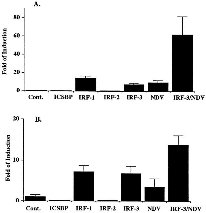
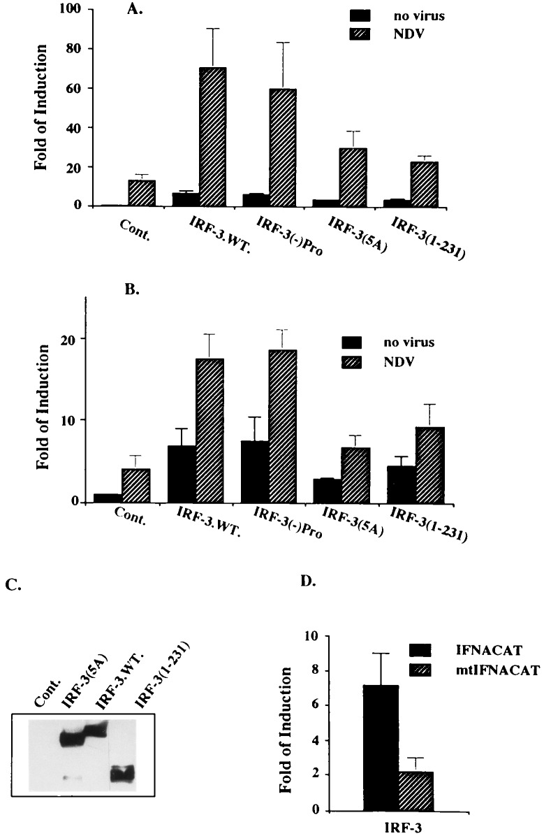
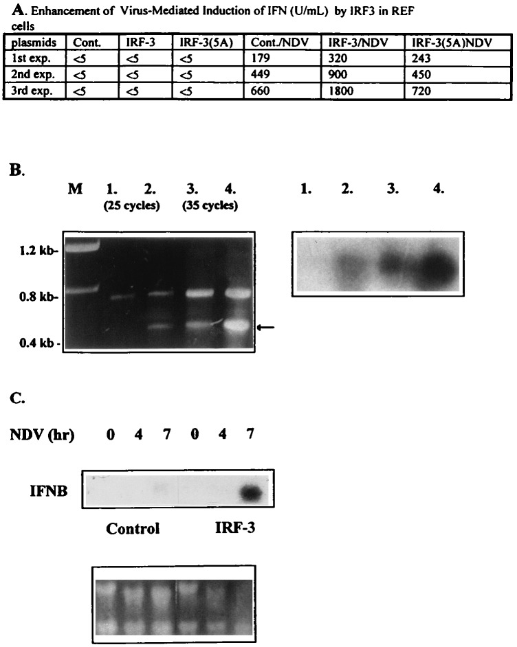
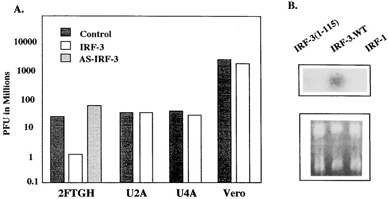
The family of interferon (IFN) regulatory factors (IRFs) encodes DNA-binding transcription factors, some of which function as modulators of virus-induced signaling. The IRF-3 gene is constitutively expressed in many tissues and cell types, and neither virus infection nor IFN treatment enhances its transcription. In infected cells, however, IRF-3 protein is phosphorylated at the carboxyl terminus, which facilitates its binding to the CBP/p300 coactivator. In the present study, we demonstrate that overexpression of IRF-3 significantly enhances virus-mediated transcription of the IFNA and IFNB genes in infected cells as well as IFN synthesis. IRF-3-mediated activation of IFN genes depends in part on carboxyl-terminal phosphorylation of a cluster of Ser/Thr residues, because a mutant with Ser/Thr to Ala substitutions activates the IFN promoter less efficiently. However, overexpression of IRF-3 in human 2FTGH cells alone results in the induction of an antiviral state, which depends on functional IFN signaling, because IRF-3 does not induce an antiviral state in mutant 2FTGH cells defective in either JAK-1 or p48 functions; also no antiviral effect of IRF-3 could be demonstrated in Vero cells that lack the IFNA and IFNB genes. This finding indicates that the observed antiviral activity of IRF-3 in 2FTGH cells results mainly from the induction of IFNs. Furthermore, E1A protein inhibited IRF-3-mediated stimulation of the IFNA4 promoter in transient expression assays; this inhibition could be reversed partially by overexpression of CBP/p300 and was not demonstrated with the mutant of E1A that does not bind p300. These results identify IRF-3 and CBP/p300 as integral components of the virus-induced complex that stimulates type 1 IFN gene transcription. The observation that adenovirus E1A antagonizes IRF-3 mediated activation suggests that E1A and IRF-3 may compete for binding to CBP/p300 and implicates a novel mechanism by which adenovirus may overcome the antiviral effects of the IFN pathway.


Figures

Figure 1
Figure 1
IRF-3 activates IFNA and IFNB promoters and cooperates with viral infection. (A) REF (rat embryonic fibroblast) cells were cotransfected with 2 μg of various IRF expressing plasmids, 2 μg of IFN4/CAT reporter plasmid, and 100 ng of pCMV-β-galactosidase as an internal control. (B) Transfection was performed as in A except 1 μg of IFNB/CAT was used. When indicated, cells were infected with NDV for 16 hr as described in Materials and Methods. CAT activity was normalized to constant levels of β-galactosidase activity. SDs are shown by T bars.
Figure 2
Figure 2
Cooperation between IRF-3 or its mutants (described in Materials and Methods) and NDV in activation of the IFNA and the IFNB promoters. The experiments were carried out as described in Fig. 1 with IFNA4/CAT reporter (A) or IFNB/CAT reporter (B). (C) Expression of IRF-3 wild type (wt) and its mutants in transfected cells used for CAT assay determined by Western blotting. (D) Two micrograms of IRF-3 was cotransfected with 2 μg of either IFNA4/CAT plasmid or mutant IFNA4/CAT plasmid (mutations are at −94 and −103 nt).
Figure 3
Figure 3
Overexpression of IRF-3 enhanced the NDV-mediated induction of endogenous IFNA4 and IFNB in REF cells. (A) Levels of biologically active IFN detected in media of infected cells. REF cells were transiently transfected with 2 μg of either pSp64 (cont.), IRF-3, or IRF-3(5A). Twenty-four hours after transfection, cells were infected with NDV for 16 hr. The media then was collected and IFN levels were determined (25). The data represent the results from three independent transfection experiments and assays. (B) RT-PCR. REF cells were transiently transfected with 4 μg of either pSp64 (lanes 1 and 3) or IRF-3 (lanes 2 and 4) by Superfect (Qiagen) transfection reagent. Two micrograms of purified RNA was amplified by RT-PCR method with specific primers for rat IFNA as described in Materials and Methods. Amplified fragments were separated on an agarose gel and visualized by ethidium bromide staining (Lt), or transferred onto nitrocellulose, and hybridized with the IFNA-specific riboprobe (Rt). (C) REF cells were transiently transfected with a control or IRF-3 plasmid. Twenty-four hours later, cells were infected for the time periods indicated. RNA was collected and analyzed by Northern hybridization to an IFNB-specific riboprobe.
Figure 4
Figure 4
Overexpression of IRF-3 activates the antiviral state in 2FTGH cells, but not in the mutants defective in IFN signaling. (A) The cells were transfected with 4 μg of IRF-3 expression plasmid and infected with VSV 16 hr posttransfection. VSV plaque assay was performed as described in the Materials and Methods. Representative experiment from three independent experiments is shown. The SD between individual experiments was less than 10%. (B) RNA was purified from 2FTGH cells transiently transfected with 4 μg of: IRF-3 (1–115), IRF-3, or IRF-1, 24 hr after transfection and analyzed by Northern blot hybridization with the IFNB-specific riboprobe.
Figure 5
Figure 5
Inhibition of IRF-3-mediated activation of the IFNA4 promoter by E1A. (A) REF cells were cotransfected with 2.0 μg of plasmids expressing IRF-1, IRF-3, or its mutants, 2 μg of IFNA4/CAT reporter plasmid, and 100 ng of pCMV-β-galactosidase in the presence and absence of E1A (2.0 μg) or E1A (Δp300) mutant. (B) The synergistic activation of IFNA promoter by IRF-3 overexpression and NDV infection did not circumvent E1A-mediated suppression. Transfection was carried out as described in A, and cells were infected 24 hr posttransfection with NDV for 16 hr as described in Materials and Methods. (C) Overexpression of p300 partially restored the IRF-3 activity from suppression by E1A. REF cells were transfected with 1.5 μg of IFNA4/CAT, 100 ng of pCMV-β-galactosidase, 1.2 μg of IRF-3 and E1A expressing plasmids, and increasing amounts (0, 0.5, 1.5 μg) of p300 expressing plasmid. (Inset) Western blotting. Expression of IRF-3 in REF cells cotransfected with IRF-3 and p300 (lanes 1–3) and E1A (lane 2) and infected with NDV (lane 3). (D) Induction of IFNA4/CAT by IRF-3 was suppressed by the cotransfection of carboxyl terminus of CBP (amino acids 1992–2441). Two micrograms of IFNA4/CAT, 100 ng of pCMV-β-galactosidase, and 1.5 μg of IRF-3 expressing plasmids were cotransfected with 0, 2, or 4 μg of Δ 5′ CBP plasmid. The CAT assay was done as described above.

References

    1. Nguyen H, Hiscott J, Pitha P M. Cyt Growth Fact Rev. 1997;8:293–312. - PubMed
    1. Harada H, Fujita T, Miyamoto M, Kimura Y, Maruyama M, Furia A, Miyata T, Taniguchi T. Cell. 1989;58:729–739. - PubMed
    1. Vaughan P S, Aziz F, van Wijnen A J, Wu S, Harada H, Taniguchi T, Soprano K J, Stein J L, Stein G S. Nature (London) 1995;377:362–365. - PubMed
    1. Lin R, Mustafa A, Nguyen H, Hiscott J. J Biol Chem. 1994;269:17542–17549. - PubMed
    1. Weisz A, Marx P, Sharf R, Appella E, Driggers P H, Ozato K, Levi B-Z. J Biol Chem. 1992;267:25589–25596. - PubMed

Publication types

MeSH terms