. 1998 Aug 18;95(17):9843-8.
doi: 10.1073/pnas.95.17.9843.

Polarity of DNA strand exchange promoted by recombination proteins of the RecA family

Affiliations

Polarity of DNA strand exchange promoted by recombination proteins of the RecA family

R C Gupta et al. Proc Natl Acad Sci U S A. .

Abstract

Homologs of Escherichia coli RecA recombination protein, which have been found throughout the living kingdom, promote homologous pairing and strand exchange. The nucleoprotein filament, within which strand exchange occurs, has been conserved through evolution, but conservation of the polarity of exchange and the significance of that directionality has not been settled. Using oligonucleotides as substrates, and assays based on fluorescence resonance energy transfer (FRET), we distinguished the biased formation of homologous joints at either end of duplex DNA from the subsequent directionality of strand exchange. As with E. coli RecA protein, the homologous Rad51 proteins from both Homo sapiens (HsRad51) and Saccharomyces cerevisiae (ScRad51) propagated DNA strand exchange preferentially in the 5' to 3' direction. The data suggest that 5' to 3' polarity is a conserved intrinsic property of recombination filaments.


Figures
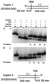
Figure 1
Figure 1
Formation of homologous joints at either end of duplex DNA in the presence of RPA. For this assay, the presynaptic filament was formed on oligonucleotide A16(−) and the duplex DNAs were duplex 1 (EG302/305) and duplex 2 (EG303/304). Duplexes 1 and 2 possessed AT-rich A16 sequences at the 3′ end or 5′ end of the displaced strand, respectively. Duplex 1 (426-mer), used for the experiment presented in A, tests for pairing at the 5′ end whereas duplex 2, a 428-mer duplex (B) detects pairing at the 3′ end. We monitored protection of the XbaI site resulting from the reaction between A16 and EG302/305 and protection of the PstI site resulting from the reaction between A16 and EG303/304. Restriction enzymes were used in an amount that was sufficient to cleave the substrate DNA in 30 sec in absence of pairing. 32P-labeled 426-mer duplex 1 (lane 1, A) and 428-mer duplex 2 (lane 1, B) were cleaved, respectively, into a 333-mer (lane 2, A) and a 335-mer (lane 2, B) fragment on restriction cleavage. When filaments were formed on a heterologous oligonucleotide, no protection of the restriction sites was observed (data not shown).
Figure 2
Figure 2
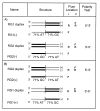
Chimeric substrates for detection of the polarity of strand exchange. The substrates used for fluorimetric assays of pairing and strand-displacement reactions. Thick and thin lines represent the GC-rich and AT-rich segments, respectively. Fluor location i and ii represent the placements of fluorescein (F) and rhodamine (R) on different strands for pairing and strand displacement, respectively. The dyes were placed at the right-hand ends of the symbolized strands: in the substrates shown in A, fluorescein was placed at the 3′ end of the single strand in the assay for pairing and at the 3′ end of the displaced strand in the assay for strand exchange; rhodamine was placed at the 5′ end of the plus strand in either assay. In B, the single-stranded oligonucleotides were the complements of those used in A, and fluorescein was located at the 5′ end of the single strand in the assay for pairing and at the 5′ end of the displaced strand in the assay for strand exchange, whereas rhodamine was at the 3′ end of the minus strand in either assay.
Figure 3
Figure 3
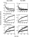
The polarity of strand exchange promoted by HsRad51, as determined by the fluorimetric assay. In AD, the substrates used were those shown in Fig. 2A. (A and B) Pairing promoted by HsRad51 without and with RPA, respectively. The labels 3′ and 5′ refer to the end of the single-stranded oligonucleotide, either RG1(−) or RG2(−), at which AT-rich sequences were located. (C and D) Strand exchange promoted by HsRad51 without and with RPA, respectively. In E and F, the substrates used were those shown in Fig. 2B in which there was an alternative arrangement of the fluorescent dyes. (E) Strand exchange in the absence of RPA. (F) Strand exchange in the presence of RPA.
Figure 4
Figure 4
The polarity of strand exchange promoted by RecA and ScRad51, as determined by the fluorimetric assay. The substrates were those shown in Fig. 2A. (A) Strand exchange by RecA. (B and C) Strand exchange by ScRad51 without and with human RPA, respectively.
Figure 5
Figure 5
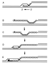
The polarity paradox. Circles indicate the location of RecA protein or one of its homologs. A spur at the end of a line designates a 3′ end. The heavy line in E represents new DNA synthesis. (A) Polarity, 5′ to 3′ with respect to the single strand on which the nucleoprotein filament had formed, would dissociate all joints. (BE) Another view of the role of polarity, according to which the polarity intrinsic to the recombination filament is only manifested in the migration of paranemic joints. Before strand invasion (B and C), 5′ to 3′ polarity would serve to drive a paranemic joint toward the 3′ end of the single strand where strand invasion (D) can occur. As in the double-strand break model for recombination, new DNA synthesis would extend and stabilize the joint (E). The subsequent action of enzymes like RuvA, B, and C can extend the region of heteroduplex DNA and finally resolve the joints into recombined products. According to this hypothesis, the polarity intrinsic to the filament only plays a biological role before strand invasion, after which the actions of other enzymes supervene.

Similar articles

Cited by

References

    1. Shinohara A, Ogawa H, Matsuda Y, Ushio N, Ikeo K, Ogawa T. Nat Genet. 1993;4:239–243. - PubMed
    1. Kowalczykowski S C, Eggleston A K. Annu Rev Biochem. 1994;63:991–1043. - PubMed
    1. Shinohara A, Ogawa H, Ogawa T. Cell. 1992;69:457–470. - PubMed
    1. Bishop D K, Park D, Xu L, Kleckner N. Cell. 1992;69:439–456. - PubMed
    1. Li M-J, Peakman M-C, Golub E I, Reddy G, Ward D C, Radding C M, Maizels N. Proc Natl Acad Sci USA. 1996;93:10222–10227. - PMC - PubMed

Publication types