. 1998 Aug 18;95(17):9885-90.
doi: 10.1073/pnas.95.17.9885.

Mechanism of capsid maturation in a double-stranded DNA virus

Affiliations

Mechanism of capsid maturation in a double-stranded DNA virus

R Tuma et al. Proc Natl Acad Sci U S A. .

Abstract

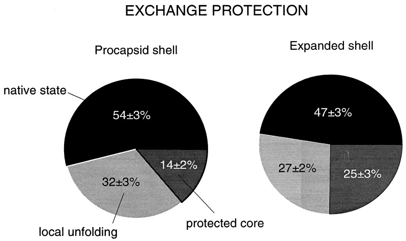
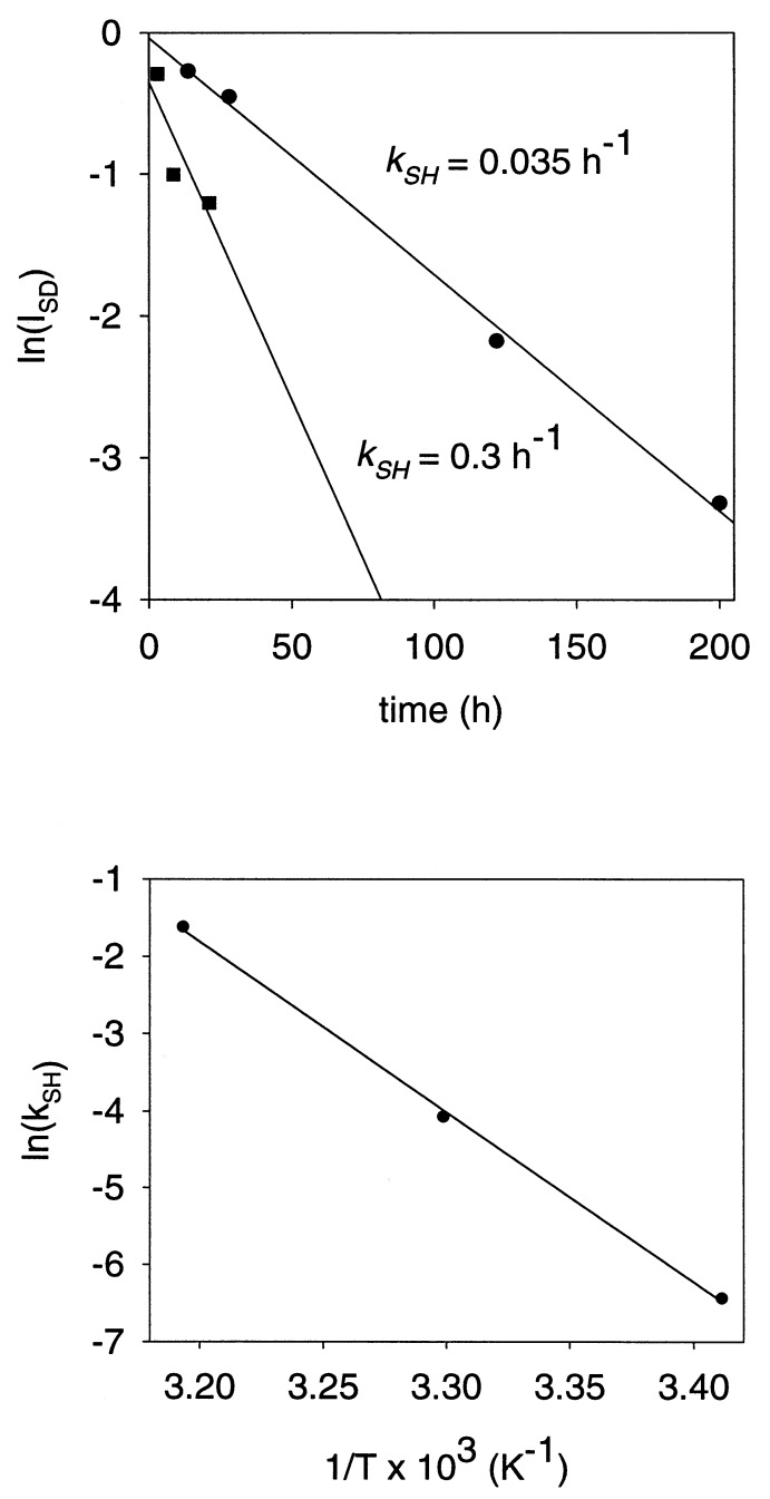
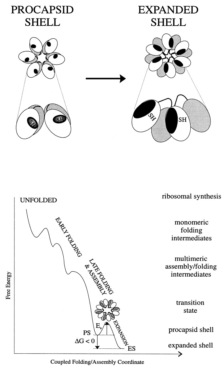
Folding mechanisms of proteins incorporated within supramolecular assemblies, including viruses, are little understood and may differ fundamentally from folding mechanisms of small globular proteins. We describe a novel Raman dynamic probe of hydrogen-isotope exchange to investigate directly these protein folding/assembly pathways. The method is applied to subunit folding in assembly intermediates of the double-stranded DNA bacteriophage P22. The icosahedral procapsid-to-capsid maturation (shell expansion) of P22 is shown to be accompanied by a large increase in exchange protection of peptide beta-strands. The molecular mechanism of shell expansion involves unfolding of metastable tertiary structure to form more stable quaternary contacts and is governed by a surprisingly high activation energy. The results demonstrate that coat subunit folding and capsid expansion are strongly coupled processes. Subunit structure in the procapsid represents a late intermediate along the folding/assembly pathway to the mature capsid. Coupling of folding and assembly is proposed as a general pathway for the construction of supramolecular complexes.


Figures

Figure 1
Figure 1
Raman monitoring of deuteration of peptide NH groups of procapsid (Upper) and expanded shells (Lower). In each panel: (A) H2O solution spectrum. (B) D2O solution spectrum of fully deuterated particle, obtained by refolding and reassembly in native D2O buffer of a sample previously treated with 6 M GuDCl. (C) Difference spectrum between A and particles exposed to D2O solution for 3 h at 10°C (signature of peptides exchanging from the native state). (D) Difference spectrum between particles exposed to D2O for 600 h at 35°C and particles exposed to D2O for 3 h at 10°C (signature of peptides exchanging via local unfolding). (E) Difference spectrum between B and particles exposed to D2O for 600 h at 35°C (signature of peptides in the exchange-protected core). Spectra were obtained from samples at 70 mg/ml concentration in 10 mM Tris buffer (pH 7.4) and 10°C.
Figure 2
Figure 2
Percentages of peptide NH groups exchanging in native, locally unfolded, and globally unfolded states of procapsid shells (54%, 32%, and 14%, respectively) and expanded shells (47%, 27%, and 25%, respectively). Exchange fractions were calculated from amide III′ band areas in the spectral interval 900-1020 cm−1. Exchange by local unfolding is defined as exchange that occurs at 35°C but not at 10°C. Exchange by global unfolding, which requires shell disassembly and subunit denaturation in 6 M GuDCl, defines peptides of the exchange-protected core.
Figure 3
Figure 3
Raman markers of the sulfhydryl group of Cys 405 in subunits of the procapsid shell (PS) and expanded shell (ES). (Right) The Raman S-H marker bands observed for H2O solutions of shells. (Left) The corresponding S-D markers observed for D2O solutions following complete SH → SD exchange.
Figure 4
Figure 4
(Upper) Rates (kSH) of Cys 405 sulfhydryl exchange in subunits of procapsid shells at 30°C (•) and expanded shells at 2°C (■). Data were obtained by measuring the intensity decay of the Raman S-H band of the shell as a function of time of exposure to D2O. (Lower) Arrhenius plot showing temperature dependence of the sulfhydryl exchange rate (kSH) in subunits of the procapsid shell. The slope of the plot corresponds to an activation energy for local unfolding (Ealocal) of 44 kcal⋅mol−1.
Figure 5
Figure 5
(Upper) Kinetics of heat-induced procapsid expansion. Procapsid shells were heated at the indicated temperatures and aliquots of the reaction mixtures were withdrawn and separated on a 1.2% (wt/vol) agarose gel. Initial expansion rates (kexpan) were estimated from gel band intensities. (Lower) Arrhenius plot showing temperature dependence of the expansion rate.
Figure 6
Figure 6
(Upper) A mechanism accounting for increased exchange protection in the expanded shell (right) vis-à-vis the procapsid shell (left). The effective increase in the subunit exchange-protected core with expansion is due to domain interchange between neighboring subunits. The shell lattice (upper diagram) is represented as a cluster of six subunits (hexon) in which the protected core is indicated by dark shading and the two coat protein domains are shown as unshaded and lightly shaded. The enlargement (lower diagram) depicts rearrangement of contacts and domains, including Cys 405 sulfhydryls, between two neighboring subunits. (Lower) Energy landscape representation of coupling between subunit folding and capsid assembly in P22. A transition state containing partially exposed hydrophobic surfaces (dark gray) is proposed for the heat-induced expansion.

Similar articles

Cited by

References

    1. Janin J. Biochimie. 1995;77:497–505. - PubMed
    1. Price N C. In: Mechanisms of Protein Folding. Pain R H, editor. New York: Oxford Univ. Press; 1994. pp. 160–193.
    1. Bai Y, Sosnick T R, Mayne L, Englander S W. Science. 1995;269:192–197. - PMC - PubMed
    1. Li T, Johnson J E, Thomas G J., Jr Biophys J. 1993;65:1963–1972. - PMC - PubMed
    1. Reilly K E, Thomas G J., Jr J Mol Biol. 1994;241:68–82. - PubMed

Publication types

LinkOut - more resources