. 1998 Aug 18;95(17):9909-13.
doi: 10.1073/pnas.95.17.9909.

A single amino acid change in the acetylcholinesterase-like domain of thyroglobulin causes congenital goiter with hypothyroidism in the cog/cog mouse: a model of human endoplasmic reticulum storage diseases

Affiliations

A single amino acid change in the acetylcholinesterase-like domain of thyroglobulin causes congenital goiter with hypothyroidism in the cog/cog mouse: a model of human endoplasmic reticulum storage diseases

P S Kim et al. Proc Natl Acad Sci U S A. .

Abstract

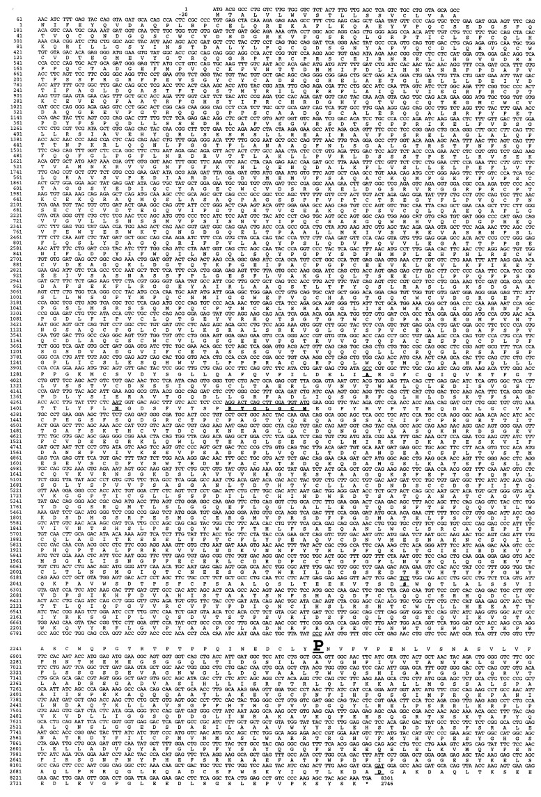
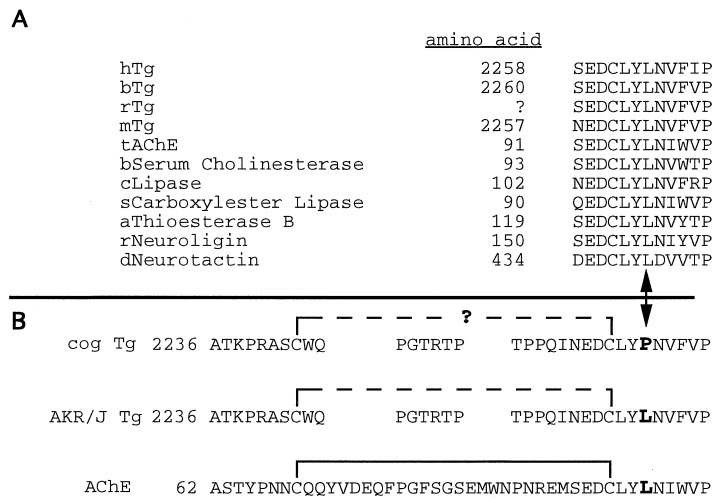
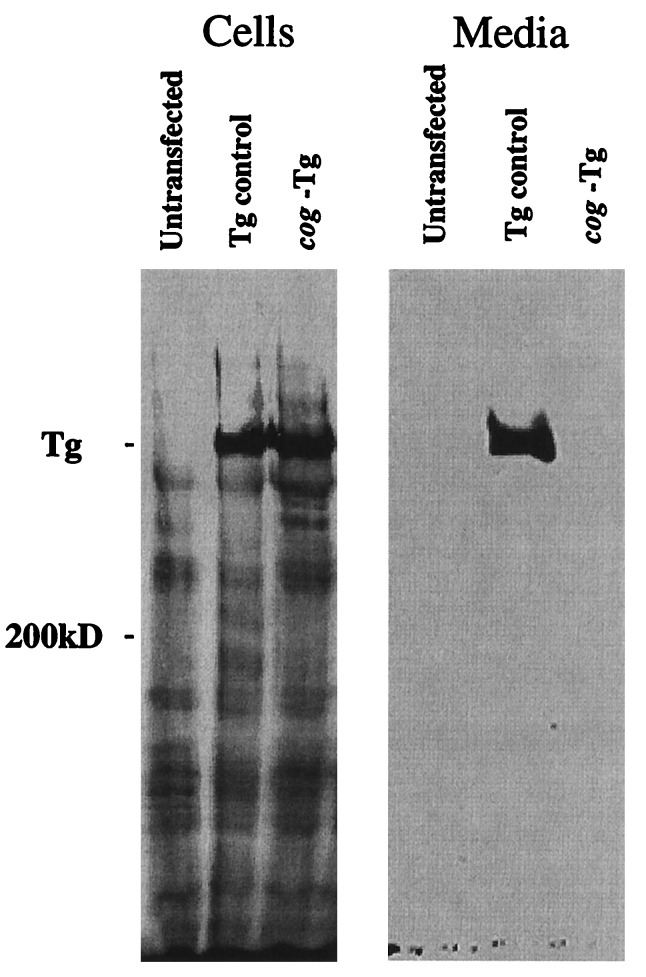
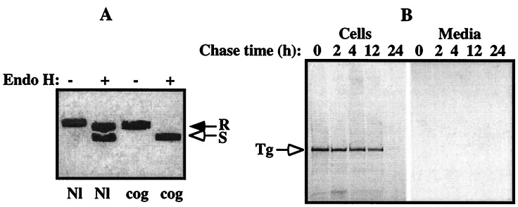
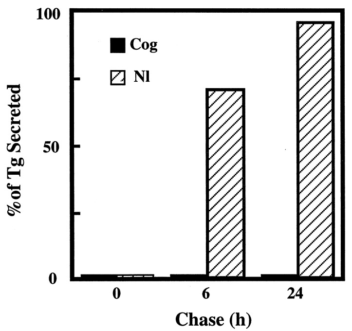
Newly synthesized thyroglobulin (Tg), the major secretory glycoprotein of the thyroid gland, folds and homodimerizes in the endoplasmic reticulum (ER) before its export to the site of iodination, where it serves as the precursor for thyroid hormone synthesis. In families with defective Tg export, affected individuals suffer from a thyroidal ER storage disease characterized by a distended thyrocyte ER containing misfolded Tg, along with induced ER molecular chaperones. Inherited as an autosomal recessive trait, deficient Tg causes congenital hypothyroidism in newborns that, if untreated, results in goiter along with serious cognitive and growth defects. Recently, a similar phenotype has been observed in inbred cog/cog mice, although the precise molecular defect has remained undefined. Here, we have isolated and cloned a full-length 8.5-kb Tg cDNA from cog/cog mice and unaffected isogenic AKR/J mice. Comparison of the complete sequences reveals that cog/cog mice express a Leu-2263 --> Pro missense mutation in the acetylcholinesterase-homology domain of Tg. Heterologous expression studies in COS cells indicate that cog Tg exhibits a severe defect in exit from the ER. Site-directed mutagenesis of cog Tg to convert the single amino acid back to Leu-2263 restores normal Tg secretion. We conclude that the cog mutation in Tg is responsible for this ER storage disease that causes thyroid dyshormonogenesis.


Figures

Figure 1
Figure 1
Tg cDNA sequence of cog/cog mice with congenital hypothyroid goiter differs by one nucleotide from that of euthyroid AKR/J mice. The complete cog Tg coding sequence and the primary structure of the deduced cog Tg protein are shown. The preparation of thyroid cDNA libraries from the two isogenic strains of mice, library screening, and DNA sequencing of selected clones, is described in Materials and Methods. The deduced cog Tg protein sequence differs in six locations from that recently reported in GenBank (accession no. U76389) for normal outbred mice (the differing codons are shown as underlined areas). Of these differences, only the nucleotide substitution (CTC to CCC) at position 6848, resulting in a Leu-2263 → Pro mutation which resides in the acetylcholinesterase-homology domain of Tg, differs between the Tg sequences of cog/cog and AKR/J mice.
Figure 2
Figure 2
The cog mutation is located within a conserved structural domain. (A) The amino acids surrounding Leu-2263 (denoted with an arrowhead, at bottom) are highly conserved in Tg from all four species that have thus far been examined, as well as in AChE and other members of the “α/β hydrolase fold” family (a partial list is shown; h, human; b, bovine; r, rat; m, mouse; t, torpedo; c, candida; s, salmon; a, avian; d, Drosophila). The amino acid numbering for rat Tg is not established because the cDNA sequence has not yet been determined in its entirety. (B) Given the close proximity of Leu-2263 to Cys-2260, which forms an intrachain disulfide bond (solid bracket) that provides important structural stability to AChE (see text) and is likely to play a similar role in Tg (dashed bracket), we hypothesize that the cog mutation (L2263P) may destabilize this region, potentially by preventing proper formation of this disulfide bridge (signified by question mark).
Figure 3
Figure 3
Defective intracellular trafficking of cog Tg in COS cells. COS-7 cells were either untransfected or transiently tranfected with Tg cDNAs as described in Materials and Methods. By Western blot analysis, unlike the control Tg, a protein encoded by the cog Tg cDNA could be found intracellularly but was secreted into the overnight collection medium at levels below the limits of detection.
Figure 4
Figure 4
Cog Tg is retained in the ER and degraded intracellularly. COS-7 cells, transfected with nonmutant or cog Tg cDNAs as in Fig. 3, were pulse-labeled with [35S]methionine/cysteine, chased for various times, and the medium collected and cells lysed as described. (A) Equal aliquots of cell lysates and chase medium were combined and immunoprecipitated with polyclonal antiserum against mouse Tg, prior to digestion with endo H (+) or mock digestion (−). As shown at 6 h of chase, ≥60% of normal Tg had passed the medial Golgi as measured by acquisition of resistance to endo H digestion (R) with secretion into the medium (not shown), whereas cog Tg remained entirely sensitive (S) to endo H digestion (open arrow), indicating that the mutant Tg protein never reached the Golgi for complex carbohydrate addition. (B) Equal aliquots of lysates of cells transfected with the cog Tg cDNA, and the corresponding chase medium samples, were immunoprecipitated with antisera against Tg and analyzed by SDS/PAGE and fluorography. Although ≈40% of cog Tg is slowly degraded over the first 12 h, during the second 12-h chase, the remaining newly synthesized cog Tg was more rapidly degraded with an undetectable fraction appearing in the secreted media, consistent with biphasic degradation kinetics (39).
Figure 5
Figure 5
Pro-2263 → Leu-corrected Tg is secreted like the wild-type protein. Using PCR-mutagenesis, the cog Tg cDNA was converted back to wild-type at residue Leu-2263. COS-7 cells, transfected with either the mutant or Pro-2263 → Leu-corrected Tg cDNA, were examined by immunoprecipitation, and SDS/PAGE as in Fig. 4. Radiolabeled Tg bands were quantitated by phosphorimaging.

Similar articles

Cited by

References

    1. Hammond C, Helenius A. Curr Opin Cell Biol. 1995;7:523–529. - PubMed
    1. Kim P S, Arvan P. Endocr Rev. 1998;19:173–202. - PubMed
    1. Ward C L, Omura S, Kopito R R. Cell. 1995;83:121–127. - PubMed
    1. Sifers R N, Finegold M J, Woo S L. Semin Liver Dis. 1992;12:301–310. - PubMed
    1. Prockop D J, Kivirikko K I. Annu Rev Biochem. 1995;64:403–434. - PubMed

Publication types

MeSH terms

Associated data