. 1998 Aug 18;95(17):9920-5.
doi: 10.1073/pnas.95.17.9920.

A putative vacuolar cargo receptor partially colocalizes with AtPEP12p on a prevacuolar compartment in Arabidopsis roots

Affiliations

A putative vacuolar cargo receptor partially colocalizes with AtPEP12p on a prevacuolar compartment in Arabidopsis roots

A A Sanderfoot et al. Proc Natl Acad Sci U S A. .

Abstract

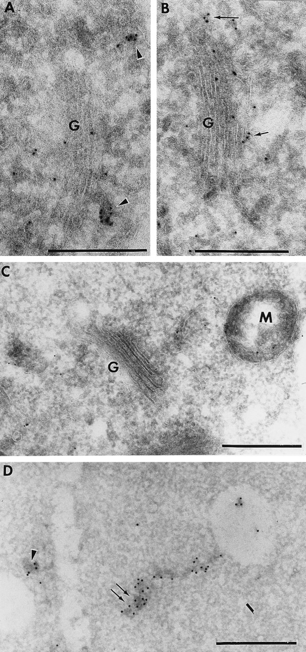
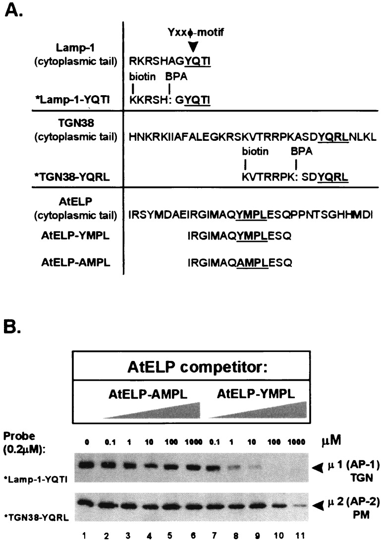
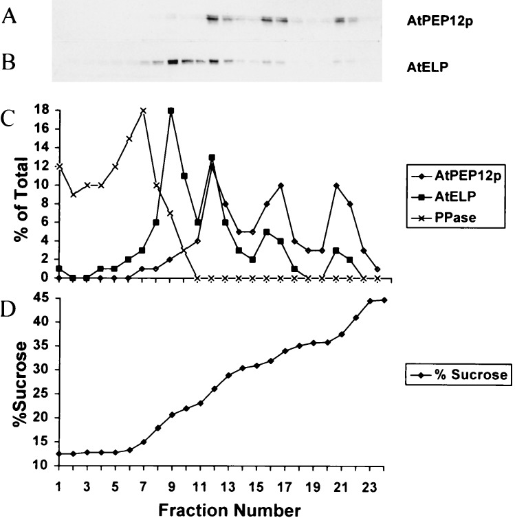
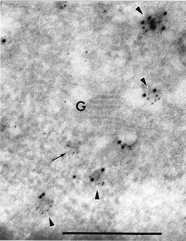
Targeting of protein cargo to the vacuole/lysosome is a multistep process that appears to have conserved features between mammalian, yeast, and plant cells. In each case, some soluble vacuolar/lysosomal proteins are believed to be bound by transmembrane cargo receptors in the trans-Golgi network (TGN) that redirect these proteins into clathrin-coated vesicles. These vesicles then appear to be transported to the prevacuole/endosome by a trafficking machinery that requires components identified in other vesicle-targeting steps such as N-ethylmaleimide-sensitive factor (NSF), soluble NSF attachment protein (SNAP), SNAP receptors (SNAREs), rab-type GTPases, and Sec1p homologs. Two likely members of this trafficking machinery have been characterized from Arabidopsis thaliana: AtPEP12p, a t-SNARE that resides on a what we now call a prevacuolar compartment, and AtELP, a protein that shares many common features with mammalian and yeast transmembrane cargo receptors. Here, we have further investigated the intracellular distribution of AtELP. We have found that AtELP is located at the trans-Golgi of Arabidopsis root cells, and that its C terminus can preferentially interact in vitro with the mammalian TGN-specific AP-1 clathrin-adapter complex, suggesting a likely role in clathrin-coated, vesicle-directed trafficking at the TGN. Further, consistent with a role in trafficking of vacuolar cargo, we have found that AtELP partially colocalizes with AtPEP12p on a prevacuolar compartment.


Figures

Figure 1
Figure 1
AtELP is localized on the trans-Golgi and post-Golgi membranes in cryosections of Arabidopsis roots. Sections were treated with antisera to AtELP (A and B), preimmune for AtELP (C), or with antisera to AtPEP12p (D) followed by biotinylated goat anti-rabbit secondary antibodies. Antibodies were then detected by streptavidin conjugated to 10 nm colloidal gold. G, Golgi; M, mitochondria. (Bar = 0.5 μm.)
Figure 2
Figure 2
Interaction of the cytoplasmic tail of AtELP with the AP-1 complex. A schematic representation of peptides used for the cross-linking studies involving AP-1 and AP-2 complexes is shown in A. Complete sequences of the Lamp-1, TGN38, and AtELP cytoplasmic tails containing the Yxxφ motif are compared with the sequences of the synthetic peptides used in this study. The Tyr-based motifs are highlighted and underlined. The *Lamp1-YQTI and *TGN38-YQRL peptides were modified by replacement of an alanine with the UV-photoactivatable, cross-linking agent benzoylphenylalanine (BPA), and by addition of biotin to the N terminus. AP-1 and AP-2 complexes (0.2 mg/ml) purified from calf brain-coated vesicles were incubated with 0.2 μM of either *Lamp1-YQTI (B Middle) or *TGN38-YQRL (B Bottom) cross-linking peptides and increasing amounts (0–1,000 μM) of the AtELP-YMPL or mutated AtELP-AMPL peptides as competitors. The appearance of the cross-linked photoreactive peptides to μ1 of AP-1 (Middle) or μ2 of AP-2 (Bottom) upon UV irradiation was tested after separation by SDS/PAGE using streptavidin conjugated to horseradish peroxidase.
Figure 3
Figure 3
Equilibrium density gradient analysis of Arabidopsis roots. Postnuclear membranes were loaded onto a sucrose step gradient and spun to equilibrium. Twenty-four fractions were collected from the top and trichloroacetic acid-precipitated, and equal volumes of each fraction were separated by SDS/PAGE and transferred to nitrocellulose. AtPEP12p (A), AtELP (B), and pyrophosphatase were detected by using specific antisera to each protein. Blots were analyzed by densitometry, and the percentage of the total marker protein detected in each fraction for AtPEP12p, AtELP, and pyrophosphatase was plotted in C. The sucrose concentration of each fraction was determined by refractometry and was plotted in D.
Figure 4
Figure 4
AtELP and AtPEP12p colocalize on a PVC in cryosections of Arabidopsis roots. Section was incubated with antisera to AtPEP12p, followed by biotinylated goat anti-rabbit secondary antibodies, then visualized with streptavidin conjugated to 10 nm colloidal gold. After a second fixation step (see Materials and Methods), the same sections were incubated with antibodies to AtELP, followed by goat anti-rabbit secondary antibodies conjugated to 5 nm colloidal gold. G, Golgi. (Bar = 0.5 μm.)

Similar articles

Cited by

References

    1. Bassham D C, Raikhel N V. Adv Bot Res. 1997;25:43–58.
    1. Robinson D G, Hinz G. Protoplasma. 1997;197:1–25.
    1. Kornfeld S. Annu Rev Biochem. 1992;61:307–330. - PubMed
    1. Marcusson E G, Hordazdovsky B F, Cereghino J L, Gharakhanian E, Emr S D. Cell. 1994;77:579–586. - PubMed
    1. Kirsch T, Paris N, Butler J M, Beevers L, Rogers J C. Proc Natl Acad Sci USA. 1994;91:3403–3407. - PMC - PubMed

Publication types

MeSH terms