. 1998 Aug 18;95(17):10038-43.
doi: 10.1073/pnas.95.17.10038.

The human GNAS1 gene is imprinted and encodes distinct paternally and biallelically expressed G proteins

Affiliations

The human GNAS1 gene is imprinted and encodes distinct paternally and biallelically expressed G proteins

B E Hayward et al. Proc Natl Acad Sci U S A. .

Abstract

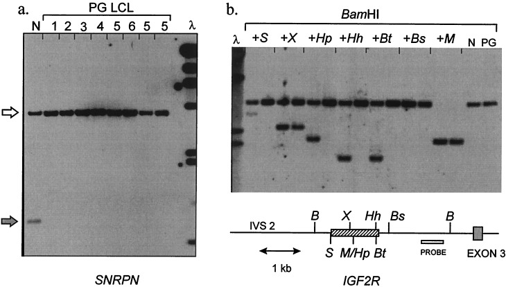
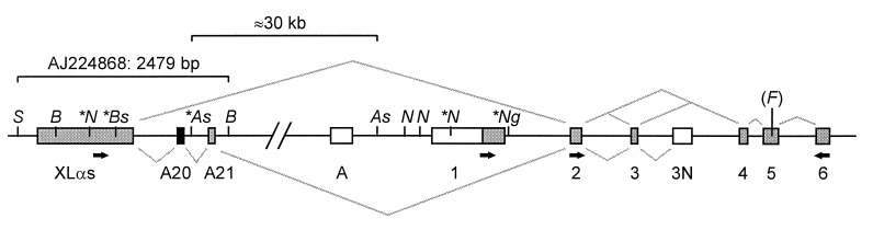
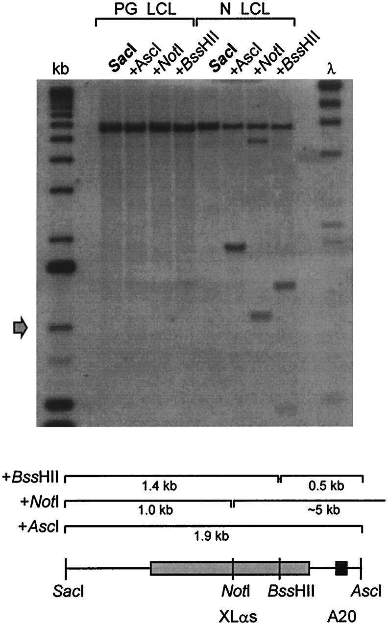
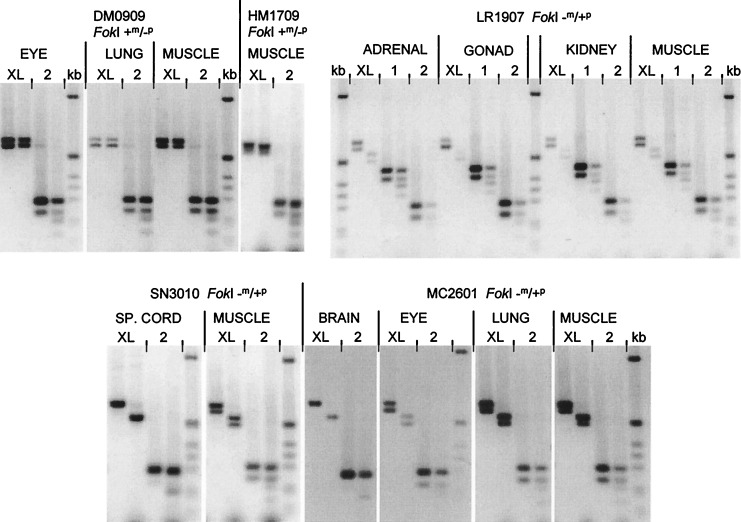
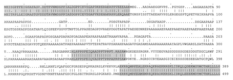
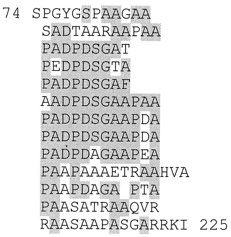
The GNAS1 gene encodes the alpha subunit of the G protein Gs, which couples receptor binding by several hormones to activation of adenylate cyclase. Null mutations of GNAS1 cause pseudohypoparathyroidism (PHP) type Ia, in which hormone resistance occurs in association with a characteristic osteodystrophy. The observation that PHP Ia almost always is inherited maternally has led to the suggestion that GNAS1 may be an imprinted gene. Here, we show that, although Gsalpha expression (directed by the promoter upstream of exon 1) is biallelic, GNAS1 is indeed imprinted in a promoter-specific fashion. We used parthenogenetic lymphocyte DNA to screen by restriction landmark genomic scanning for loci showing differential methylation between paternal and maternal alleles. This screen identified a region that was found to be methylated exclusively on a maternal allele and was located approximately 35 kb upstream of GNAS1 exon 1. This region contains three novel exons that are spliced into alternative GNAS1 mRNA species, including one exon that encodes the human homologue of the large G protein XLalphas. Transcription of these novel mRNAs is exclusively from the paternal allele in all tissues examined. The differential imprinting of separate protein products of GNAS1 therefore may contribute to the anomalous inheritance of PHP Ia.


Figures

Figure 1
Figure 1
Uniparental methylation pattern at imprinted loci in human parthenogenetic LCL. (a) Methylation of SNRPN (chromosome 15q11-q13). The probe used for Southern blotting is KB17, a standard diagnostic reagent for Prader–Willi syndrome. DNAs are digested with XbaI + NotI. The paternal allele (gray arrow) is unmethylated, yielding a 0.9-kb fragment from normal DNA (N). 1–6, six subcultured lines of parthenogenetic lymphoblastoid cells (LCL) established from patient F.D. Only the methylated 4.2-kb maternal band (white arrow) is present. λ, λHindIII marker; 23, 9.4, 6.6, 4.4, 2.3, 2.0, 0.56 kb. (b) Methylation of IGF2R. The probe (open box) is fragment Bx of clone pE3UP (25), which detects a 3.2-kb BamHI fragment. Pairs of lanes contain normal (N) or parthenogenetic (PG) LCL DNA digested with BamHI, alone or plus a second enzyme as indicated: B, BamHI; S, SacII; X, XhoI; M/Hp, MspI/HpaII; Hh, HhaI; Bt, BstUI; Bs, BssHII. For each enzyme only the site nearest the probe is shown. The cross-hatched box indicates the region of allele-specific methylation; the HpaII, HhaI, and BstUI sites are methylated only on a maternal allele; the SacII site in addition is methylated incompletely on a paternal allele; the XhoI is methylated incompletely on a maternal allele and unmethylated on a paternal allele. These results concur with previous data (25) and indicate faithful allele-specific methylation across this region in the PG LCL DNA. λ, λHindIII marker; 6.6, 4.4, 2.3, 2.0 kb.
Figure 2
Figure 2
Organization of the XLαs region of GNAS1. The map is not to scale, and not all restriction sites are shown, with the exception of AscI and NotI. Grey lines above the exons represent the splicing patterns observed in the majority of XLαs-containing RT-PCR products and below the line the splicing pattern seen in the 359933 cDNA clone. Italic letters indicate restriction sites: S, SacI; B, BamHI; N, NotI; As, AscI; Ng, NgoMI; Bs, BssHII; (F), FokI (polymorphic site). Asterisks indicate those sites whose methylation status was assessed by Southern blotting (see text). The distance between the two AscI sites is ≈30 kb. The 2.5-kb genomic region that was sequenced is indicated. The A20 exon is black; other exons are gray or white for untranslated regions. The exon labeled A has been described (26). Small black arrows indicate approximate positions of primers used for RT-PCR.
Figure 3
Figure 3
Differential methylation in the XLαs region of GNAS1. The probe used was a 1.9-kb SacI–AscI genomic fragment containing both the XLαs and A20 exons (shaded boxes). Genomic DNA from parthenogenetic (left) or normal (right) lymphoblastoid cell DNA was digested with SacI, alone or in combination with AscI, NotI, or BssHII. SacI generates a fragment of ≈6 kb; the expected fragment sizes derived from additional cleavage at the methylation sensitive sites are indicated on the diagram. λ, λHindIII marker; 23, 9.4, 6.6, 4.4, 2.3, 2.0, 0.56 kb. kb, 1-kb ladder (GIBCO/BRL); upwards from arrow 1.0, 1.6, 2.0, 3.1, 4.1, 5.1, 6.1, 7.1 kb.
Figure 4
Figure 4
Exclusively paternal expression of XLαs-containing GNAS1 transcripts. All experiments used a downstream PCR primer in exon 6; the location of the upstream primer is indicated (either in the XLαs exon, exon 1 or exon 2) above each pair of lanes. Each pair of lanes contains undigested (Left) or FokI-digested (Right) RT-PCR product. The two bands seen in most undigested samples result from alternative splicing of exon 3. In fetuses DM0909 and HM1709, the paternal allele is lacking a FokI site (genotype +m/−p). The XLαs-derived RT-PCR products (XL) are completely FokI-resistant. The total GNAS1 transcripts, in contrast (2), are partially cleaved by FokI, indicating their biallelic origin. In fetus LR1907, the genotype is −m/+p. The XLαs-derived products are digested to completion by FokI, again demonstrating their exclusively paternal origin. Also, in this panel, exon 1-specific transcription has been analyzed separately, showing biallelic expression from the promoter upstream of exon 1. Fetuses SN3010 and MC2601 are again FokI −m/+p. RT-PCR products are digested to completion if XLαs-derived but only partly when the exon 2 primer is used. kb, 1-kb ladder: 1,018, 517/506, 396, 344, 298, 220, 201, 154, 134 bp.
Figure 5
Figure 5
Comparison between human (Upper) and rat (Lower) XLαs peptide sequences. Extensive homology is confined to the three shaded regions, whereas, as discussed in the text, the central repetitive region aligns poorly between species. Translation of the human exon has been started at position 318 relative to the SacI site (AJ224868). However, the true translation initiation site has not been defined experimentally either for human or rat XLαs (8).
Figure 6
Figure 6
Internal repeat motifs in the human XLαs exon. Almost the whole of the central poorly conserved region is included in this repeat, which begins at residue 74 and ends at residue 225, immediately before the second highly conserved region (shaded in Fig. 5).

Similar articles

Cited by

References

    1. Kozasa T, Itoh H, Tsukamoto T, Kaziro Y. Proc Natl Acad Sci USA. 1988;85:2081–2085. - PMC - PubMed
    1. Levine M A, Ahn T G, Klupt S F, Kaufman K D, Smallwood P M, Bourne H R, Sullivan K A, Van Dop C. Proc Natl Acad Sci USA. 1988;85:617–621. - PMC - PubMed
    1. Patten J L, Johns D R, Valle D, Eil C, Grupposo P A, Steele G, Smallwood P M, Levine M A. N Engl J Med. 1990;322:1412–1419. - PubMed
    1. Weinstein L S, Gejman P V, Collins R M, Gershon E S, Spiegel A M. Proc Natl Acad Sci USA. 1990;87:8287–8290. - PMC - PubMed
    1. Davies S J, Hughes H E. J Med Genet. 1993;30:101–103. - PMC - PubMed

Publication types

MeSH terms

Substances

Associated data