

Widespread expression of an autoantigen-GAD65 transgene does not tolerize non-obese diabetic mice and can exacerbate disease
- PMID: 9707599
- PMCID: PMC21460
- DOI: 10.1073/pnas.95.17.10055
Widespread expression of an autoantigen-GAD65 transgene does not tolerize non-obese diabetic mice and can exacerbate disease
Abstract
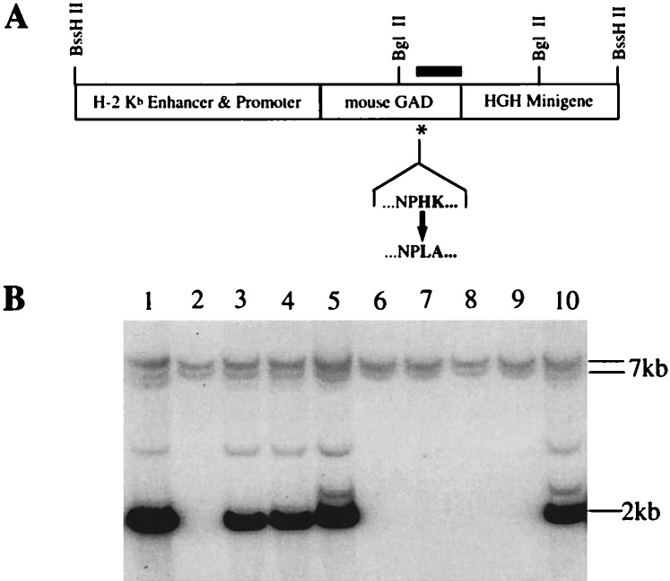
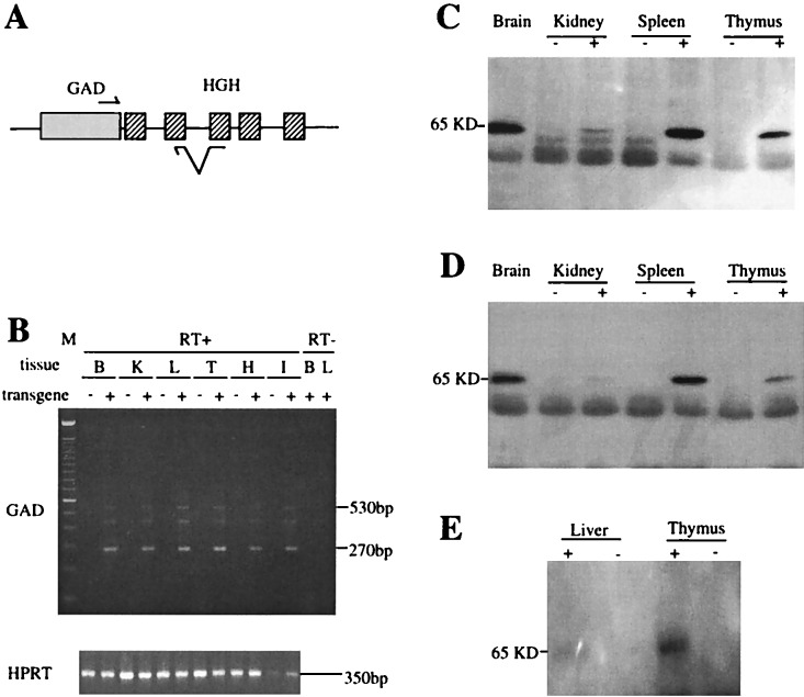
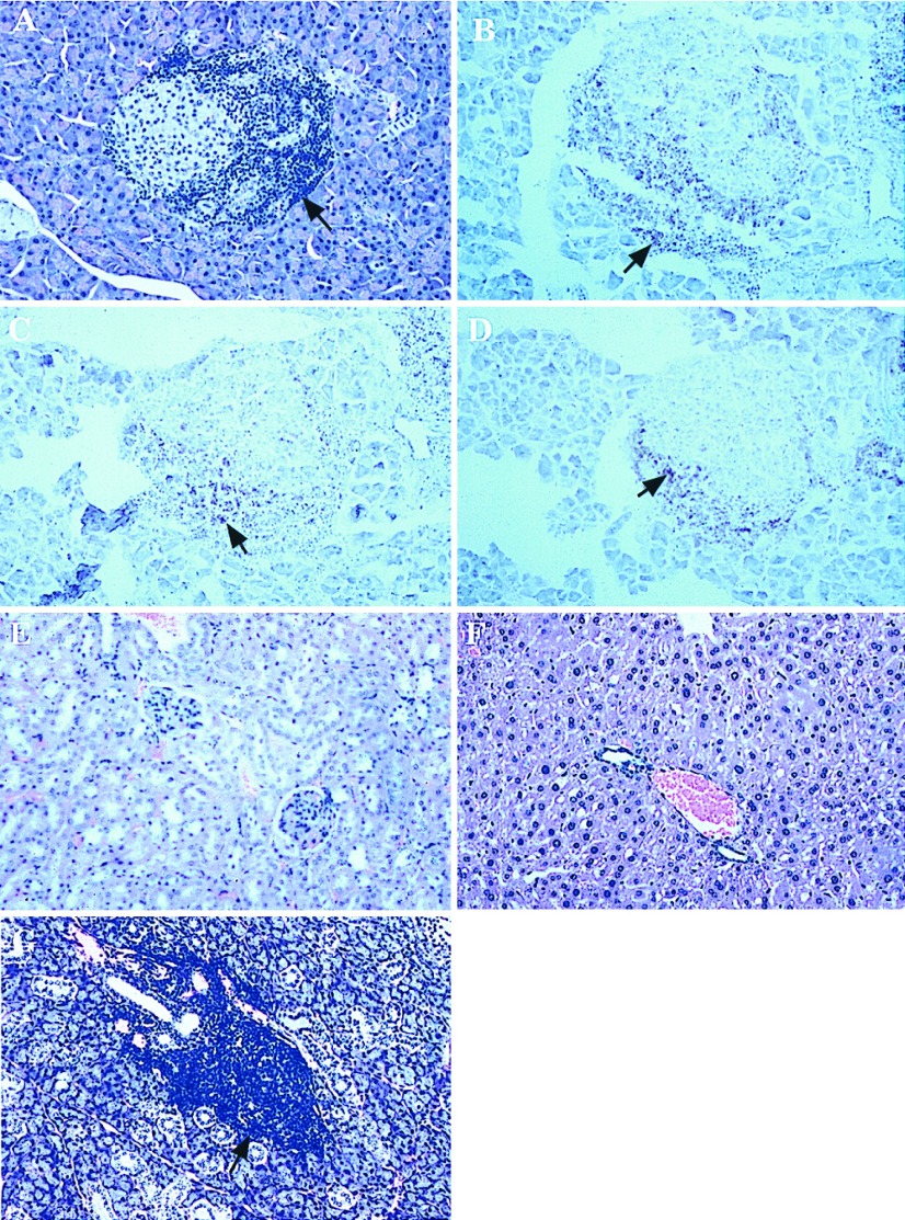
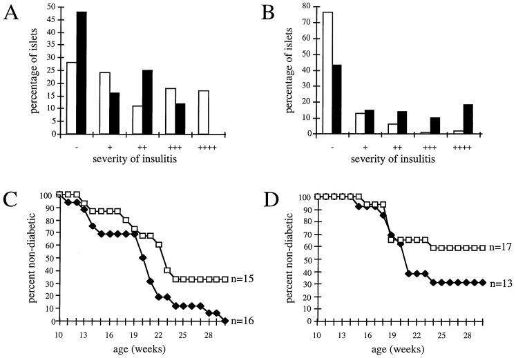
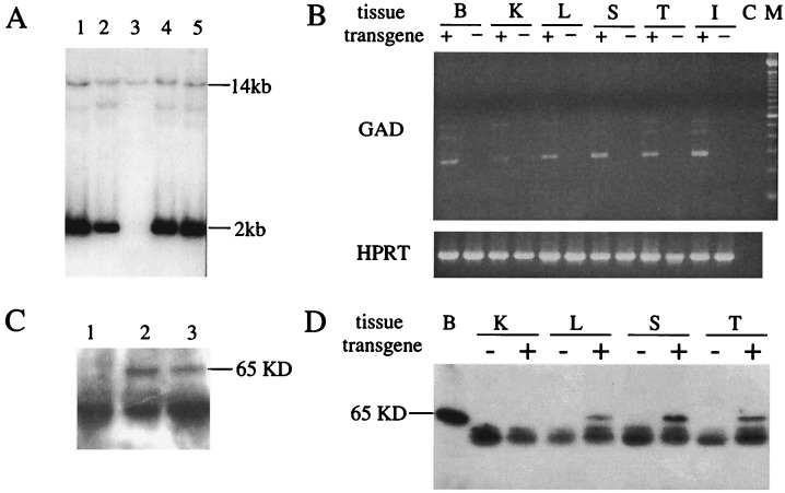
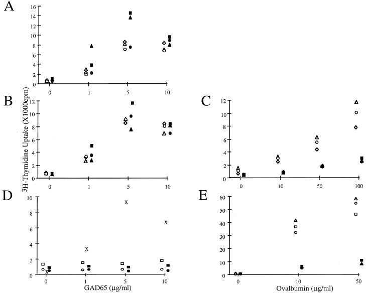
Glutamic acid decarboxylase (GAD)65 is a pancreatic beta cell autoantigen implicated as a target of T cells that initiate and sustain insulin-dependent diabetes mellitus (IDDM) in humans and in non-obese diabetic (NOD) mice. In an attempt to establish immunological tolerance toward GAD65 in NOD mice, and thereby to test the importance of GAD in IDDM, we generated three lines transgenic for murine GAD65 driven by a major histocompatibility complex class I promoter. However, despite widespread transgene expression in both newborn and adult mice, T cell tolerance was not induced. Mononuclear cell infiltration of the islets (insulitis) and diabetes were at least as bad in transgenic mice as in nontransgenic NOD mice, and in mice with the highest level of GAD65 expression, disease was exacerbated. In contrast, the same transgene introduced into mouse strain, FvB, induced neither insulitis nor diabetes, and T cells were tolerant to GAD. Thus, the failure of NOD mice to develop tolerance toward GAD65 reflects at minimum a basic defect in central tolerance, not seen in animals not predisposed to IDDM. Hence, it may not be possible experimentally to induce full tolerance toward GAD65 in prediabetic individuals. Additionally, the fact that autoimmune infiltration in GAD65 transgenic NOD mice remained largely restricted to the pancreas, indicates that the organ-specificity of autoimmune disease is dictated by tissue-specific factors in addition to those directing autoantigen expression.
Figures
Similar articles
-
Differential protection in two transgenic lines of NOD/Lt mice hyperexpressing the autoantigen GAD65 in pancreatic beta-cells.Diabetes. 1998 Dec;47(12):1848-56. doi: 10.2337/diabetes.47.12.1848. Diabetes. 1998. PMID: 9836515
-
Major DQ8-restricted T-cell epitopes for human GAD65 mapped using human CD4, DQA1*0301, DQB1*0302 transgenic IA(null) NOD mice.Diabetes. 1999 Mar;48(3):469-77. doi: 10.2337/diabetes.48.3.469. Diabetes. 1999. PMID: 10078545
-
Immune response to glutamic acid decarboxylase correlates with insulitis in non-obese diabetic mice.Nature. 1993 Nov 4;366(6450):72-5. doi: 10.1038/366072a0. Nature. 1993. PMID: 8232539
-
Genetic and immunological basis of autoimmune diabetes in the NOD mouse.Rev Immunogenet. 2000;2(1):140-6. Rev Immunogenet. 2000. PMID: 11324686 Review.
-
Cellular and molecular roles of beta cell autoantigens, macrophages and T cells in the pathogenesis of autoimmune diabetes.Arch Pharm Res. 1999 Oct;22(5):437-47. doi: 10.1007/BF02979150. Arch Pharm Res. 1999. PMID: 10549569 Review.
Cited by
-
Dissecting autoimmune diabetes through genetic manipulation of non-obese diabetic mice.Diabetologia. 2003 Nov;46(11):1447-64. doi: 10.1007/s00125-003-1218-1. Epub 2003 Oct 28. Diabetologia. 2003. PMID: 14586501 Review.
-
Regulation of GAD65 expression by SMAR1 and p53 upon Streptozotocin treatment.BMC Mol Biol. 2012 Sep 14;13:28. doi: 10.1186/1471-2199-13-28. BMC Mol Biol. 2012. PMID: 22978699 Free PMC article.
-
Prevention of type I diabetes transfer by glutamic acid decarboxylase 65 peptide 206-220-specific T cells.Proc Natl Acad Sci U S A. 2004 Sep 28;101(39):14204-9. doi: 10.1073/pnas.0405500101. Epub 2004 Sep 20. Proc Natl Acad Sci U S A. 2004. PMID: 15381770 Free PMC article.
-
Transgenic models of autoimmune disease.Clin Exp Immunol. 2002 Jan;127(1):4-11. doi: 10.1046/j.1365-2249.2002.01771.x. Clin Exp Immunol. 2002. PMID: 11882026 Free PMC article. Review.
-
Type 1 diabetes pathogenesis and the role of inhibitory receptors in islet tolerance.Ann N Y Acad Sci. 2020 Feb;1461(1):73-103. doi: 10.1111/nyas.14106. Epub 2019 Apr 26. Ann N Y Acad Sci. 2020. PMID: 31025378 Free PMC article. Review.
References
Publication types
MeSH terms
Substances
Grants and funding
LinkOut - more resources
Full Text Sources
Other Literature Sources
Medical
Molecular Biology Databases
Research Materials
