. 1998 Aug 18;95(17):10188-93.
doi: 10.1073/pnas.95.17.10188.

Hypoxia stimulates insulin-like growth factor binding protein 1 (IGFBP-1) gene expression in HepG2 cells: a possible model for IGFBP-1 expression in fetal hypoxia

Affiliations

Hypoxia stimulates insulin-like growth factor binding protein 1 (IGFBP-1) gene expression in HepG2 cells: a possible model for IGFBP-1 expression in fetal hypoxia

S I Tazuke et al. Proc Natl Acad Sci U S A. .

Abstract

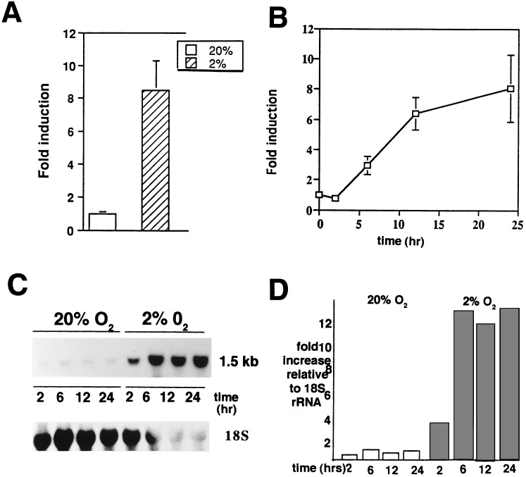
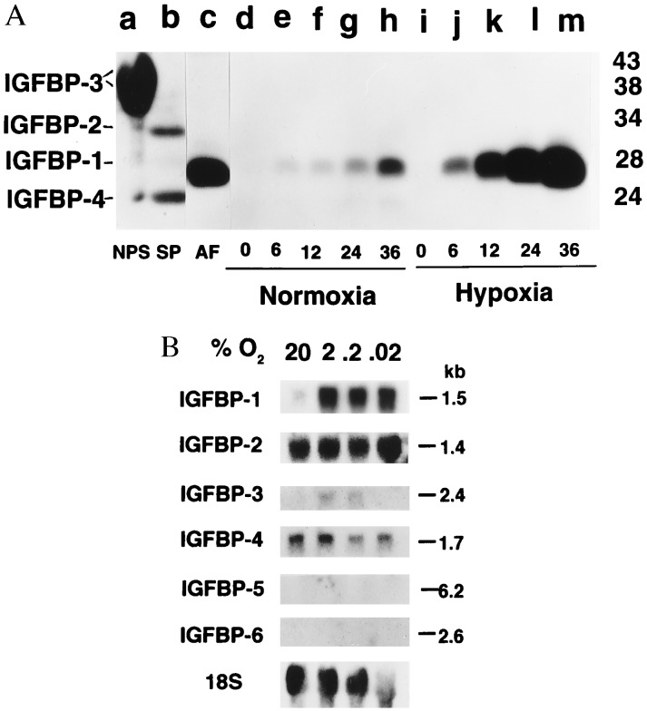
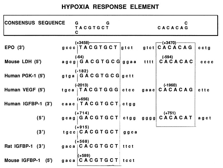
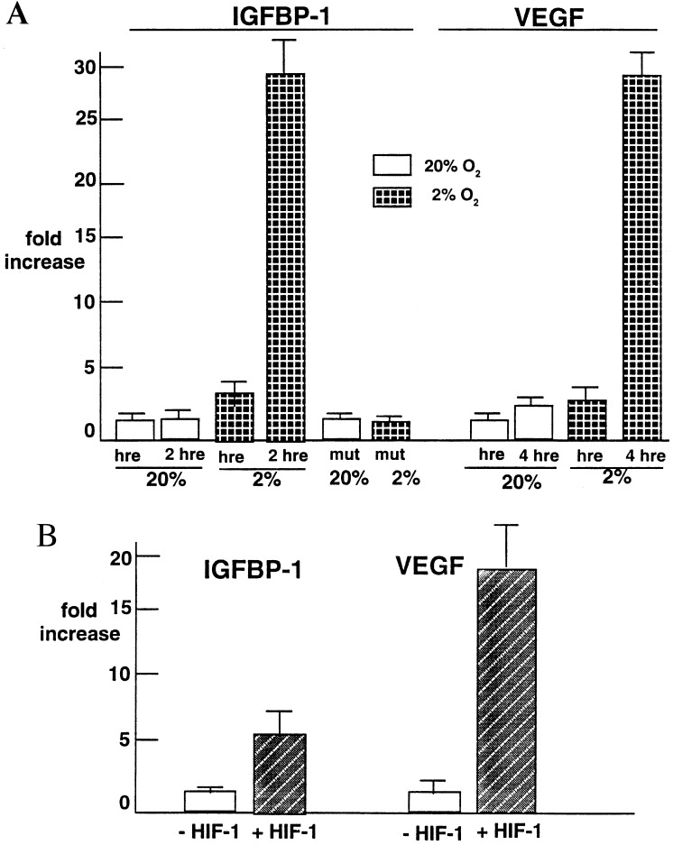
IGFBP-1 is elevated in fetuses with long-term, chronic hypoxia and intrauterine growth restriction. We investigated the hypothesis that hypoxia regulates IGFBP-1 in the human fetus in vivo and IGFBP-1 gene expression and protein in vitro. Umbilical artery IGFBP-1 levels (mean +/- SEM) from term babies with respiratory acidosis (acute hypoxia), normal babies, and those with mixed respiratory/metabolic acidosis (more profound and prolonged hypoxia) were measured using an immunoradiometric assay. IGFBP-1 levels were similar in normal (n = 12) and acutely hypoxic (n = 6) babies (189.1 +/- 71.8 vs. 175.8 +/- 45.9 ng /ml, respectively, P = 0.789). However, with more profound and prolonged hypoxia (n = 19), IGFBP-1 levels were markedly elevated (470.6 +/- 80.0 ng /ml, P = 0.044). To investigate IGFBP-1 regulation by hypoxia in vitro, HepG2 cells were incubated under hypoxia (pO2 = 2%) and normoxia (pO2 = 20%). IGFBP-1 protein and mRNA increased 8- and 12-fold, respectively, under hypoxic conditions. Hypoxia did not affect protein or mRNA levels of IGFBP-2 or -4. IGFBP-5 and -6 mRNAs, undetectable in control cells, were not induced by hypoxia, whereas minimally expressed IGFBP-3 mRNA increased twofold. Investigation into IGFBP-1 gene structure revealed three potential consensus sequences for the hypoxia response element (HRE) in the first intron. To investigate functionality, a 372-bp fragment of IGFBP-1 intron 1, containing putative HREs, was placed 5' to a heterologous hsp70 promoter in a plasmid using luciferase as a reporter gene. Under hypoxia, reporter gene activity increased up to 30-fold. Mutations in the middle HRE abolished reporter activity in response to hypoxia, suggesting that this HRE is functional in the IGFBP-1 hypoxia response. Cotransfection of HRE reporter genes with a constitutively expressing hypoxia-inducible factor 1 plasmid in HepG2 cells resulted in a fourfold induction of reporter activity, suggesting a role for hypoxia-inducible factor 1 in hypoxia induction of IGFBP-1 gene expression. These data support the hypothesis that hypoxia regulation of IGFBP-1 may be a mechanism operating in the human fetus to restrict insulin-like growth factor-mediated growth in utero under conditions of chronic hypoxia and limited substrate availability.


Figures

Figure 1
Figure 1
Hypoxia induction of IGFBP-1 in HepG2 cells. (A) Cells were grown in serum-containing medium in the presence of 20% and 2% O2, respectively. After 24 hr, the conditioned medium was collected and assayed in triplicate for IGFBP-1 using an immunoradiometric assay. Values shown are the mean ± SEM of six different experiments. (B) Time course of hypoxia experiments, with media being harvested after 2, 6, 12, and 24 hr of exposure of HepG2 cells at 2% and 20% pO2. Data represent the mean of four experiments run in triplicate and are reported as “fold induction.” (C) At the end of each time period, total RNA was collected from the cells and a 1.5-kb mRNA transcript was detected by Northern blot analysis using an EcoRI fragment (938 bp) of the IGFBP-1 cDNA. 18S rRNA shown at the bottom. (D) Densitometry of Northern blot shown in C, with fold increase normalized to 18S rRNA.
Figure 2
Figure 2
Specificity of IGF-binding protein response to hypoxia. (A) Autoradiogram of representative Western ligand blot of media conditioned by HepG2 cells cultured under normoxia (20% O2, lanes d–h) and hypoxia (2% O2, lanes i–m) for time (in hr) shown. Controls are in lanes a–c. Nonpregnant human serum (NPS, lane a) demonstrates IGFBP-3, a prominent doublet with relative molecular mass between 38 and 43 kDa. Lane b shows IGFBP-2 (34 kDa) and IGFBP-4 (24 kDa) in human seminal plasma (SP). Human midgestation amniotic fluid (AF, lane c) shows IGFBP-1 (28 kDa). Molecular mass markers are shown on the right in kDa and migration of IGFBPs is shown on the left. (B) Autoradiogram of representative Northern blot of total RNA isolated from HepG2 cells cultured 24 hr in normoxia (20%) or hypoxia at % O2 shown. Membrane was probed sequentially with 32P-labeled cDNA probes (see text and Materials and Methods). Transcript sizes are shown on the right in kb. Exposure times were: for IGFBP-1, 2 hr; IGFBP-2, 3 days; IGFBP-3, 30 days; IGFBP-4, 3 days; IGFBP-5 and IGFBP-6, 7 days; 18S, 16 hr.
Figure 3
Figure 3
Comparison of the HRE consensus sequences and CACACAG/C elements in proximal intron 1 of human, rat, and mouse IGFBP-1 genes and select hypoxia-regulated genes. Nucleotide numbers are those used in GenBank sequences [human (37), rat (42), mouse (43)]. The putative HREs are shown in uppercase letters and are encased by a box. LDH, lactate dehydrogenase; PGK, phosphoglycerokinase.
Figure 4
Figure 4
HRE and HIF-1 involvement in IGFBP-1 response to hypoxia. (A) Induction of reporter gene activity in HepG2 cells by hypoxia (expressed as fold increase above control, see text). Shown are the mean ± SD for four different experiments for IGFBP-1 HRE constructs and three different experiments with VEGF HRE constructs. Two-way analysis of variance revealed significantly increased reporter gene activity with a single copy of the IGFBP-1 HRE in the construct (P = 0.02) and with two copies of it (P = 0.0001). In normoxia, two copies of the IGFBP-1 HRE construct did not result in an increase in luciferase activity. hre, hypoxia response element; mut, mutant. (B) Transient transfection studies with the HRE in intron 1 of the IGFBP-1 gene attached to a luciferase reporter gene in a reporter construct cotransfected with a constitutively expressing HIF-1α plasmid (+HIF-1) or without (−HIF-1). Fold increase in luciferase activity is shown. VEGF served as a control (see text). Results show the mean ± SD of three different experiments. Two-way ANOVA revealed significantly increased reporter gene activity with cotransfection with the HIF-1α plasmid compared with controls (P = 0.001) and with two copies (P = 0.0001).

Similar articles

Cited by

References

    1. Creasy R K, Resnik R. In: Maternal-Fetal Medicine: Principles and Practice. Creasy R K, Resnik R, editors. Philadelphia: Saunders; 1993. pp. 547–563.
    1. Bang P, Giudice L C, Rosenfeld R G. Front Endocrinol. 1994;6:197–212.
    1. Chard T. Growth Regul. 1994;4:91–100. - PubMed
    1. Baker J, Liu J P, Robertson E J, Efstratiadis A. Cell. 1993;75:73–82. - PubMed
    1. Woods K A, Camacho-Hubner C, Savage M O, Clark A J L. N Engl J Med. 1996;335:1363–67. - PubMed

Publication types

MeSH terms

Substances