. 1998 Aug 18;95(17):10218-23.
doi: 10.1073/pnas.95.17.10218.

Propagation by sporulation in the guinea pig symbiont Metabacterium polyspora

Affiliations

Propagation by sporulation in the guinea pig symbiont Metabacterium polyspora

E R Angert et al. Proc Natl Acad Sci U S A. .

Abstract

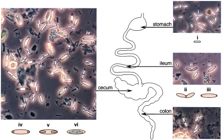
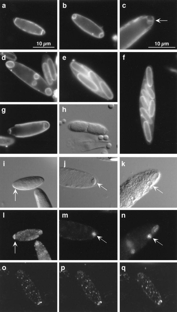
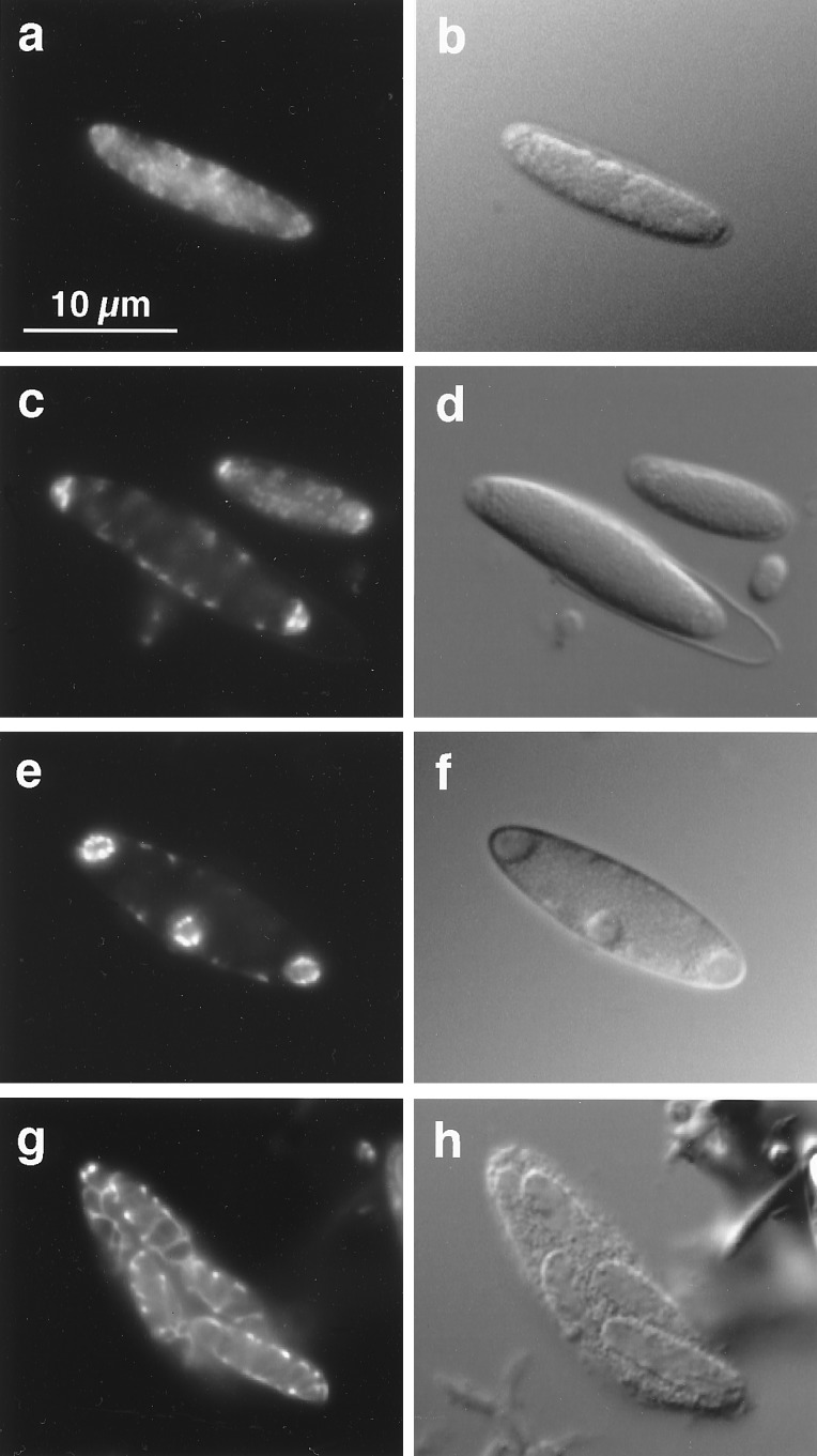
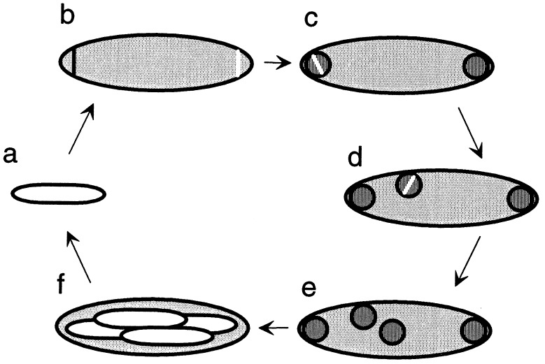
The Gram-positive bacterium Metabacterium polyspora is an uncultivated symbiont of the guinea pig gastrointestinal tract. Here we present evidence that in M. polyspora vegetative cell division has taken on a minor, and apparently dispensable, role in propagation. Instead, this unusual bacterium has evolved the capacity to produce progeny in the form of multiple endospores. Endospore formation is coordinated with transit of the bacterium through the gastrointestinal tract of the guinea pig. For the majority of cells, sporulation is initiated in the ileum, whereas later stages of development take place in the cecum. We show that multiple endospores are generated both by asymmetric division at both poles of the cell and by symmetric division of the endospores at an early stage of their development. Our findings suggest that M. polyspora represents an intermediate step in the evolution of a novel mode of cellular propagation that originates with endospore-forming Bacillus and Clostridium spp., which reproduce by binary fission, and extends to Epulopiscium spp., which create multiple viviparous offspring by a process of internal reproduction.


Figures

Figure 1
Figure 1
The life cycle of M. polyspora is coordinated with its passage through the gastrointestinal tract of the guinea pig (Cavia porcellus). The stomach contains only free, mature endospores (i). The ileum contains cells undergoing binary fission (ii) and cells with asymmetrically positioned septa (iii). [Cells undergoing binary fission (ii) are surrounded by a spore coat, which is best seen under higher magnification and by DIC microscopy as in Fig. 2h. Likewise, the presence of polar septa in cells (iii) is more clearly seen at higher magnification and by use of a fluorescent membrane stain as in Fig. 2a.] The majority of cells in the cecum contain either developing forespores, seen as phase-dark bodies within the cell (iv and v) or phase-bright, mature endospores (vi). Panels show phase-contrast micrographs of samples of contents taken from the organ indicated.
Figure 2
Figure 2
Sporulation, binary fission, and FtsZ localization. ae show a series of cells illustrating successive stages of sporulation in M. polyspora. (a) This cell contains two asymmetrically positioned septa, indicative of the start of sporulation. (b) The phagocytic process of forespore engulfment can be seen in this cell. (c) The arrow indicates a membrane septum that has divided a single forespore protoplast in two. (d) Four immature forespores are visible within this sporangium. (e) The outline of four mature endospores can be seen within this sporangium. (f) A large cell with seven visible endospores. (g) In this germinating spore, asymmetrically positioned septa are visible even as the cell emerges from its spore coat. (h) This DIC micrograph shows cells just after binary fission and still contained within a single spore coat. Cells (a–g) were treated with a fluorescent membrane stain, either FM 4–64 or FM 1–43 (which we found also have an affinity for spore coats). iq summarize the results of the FtsZ localization experiments. Pairs of panels show cells of the same field viewed with DIC (i–k) and fluorescence (l–n) microscopy. Arrows indicate the position of the FtsZ ring. In i and l, FtsZ is localized to the cell pole. j and m show an FtsZ ring bisecting a newly engulfed forespore. k and n show a forespore at a more lateral position undergoing symmetric division. (oq) 0°, 15°, and 30° vertical plane rotations, respectively, of a three-dimensional reconstruction of a cell with FtsZ localized in a ring-like structure near one cell pole. All panels except d are represented at the same magnification. The scale bar for these images is shown in a.
Figure 3
Figure 3
Distribution of DNA during sporulation in M. polyspora as seen in cells stained with the DNA-specific dye 4′,6-diamidino-2-phenylindole (DAPI). This figure shows pairs of micrographs of the same cell with 4′,6-diamidino-2-phenylindole (DAPI) fluorescence on the left and DIC on the right. (a and b) Prior to septation DNA is seen dispersed throughout the cell. (c and d) After septation highly condensed DNA is found in each forespore while DNA remaining in the mother cell is arranged about the cell periphery. This pattern of DNA distribution is similar to that observed in Epulopiscium sp. early in daughter cell development (23). Note that the next round of sporulation has been initiated in the larger cell shown here even before it has emerged completely from its spore coat. (e and f) As forespores enlarge the DNA takes on a peripheral arrangement. (g and h) In large forespores the DNA assumes a highly ordered pattern.
Figure 4
Figure 4
Sporulation and the life cycle of M. polyspora. As an endospore (a) germinates, the emerging bacterium often shows signs of the initiation of the next round of sporulation. (b) Two forespores are formed by the production of two asymmetrically positioned septa, one near each cell pole. This septation is mediated by the cell division protein FtsZ, shown here as a white line spanning a membrane-bound compartment (see also Fig. 2). (c) A newly engulfed forespore may undergo fission. (d) Forespore fission can also occur later, after a forespore has migrated to a lateral position within the mother cell. (e) The forespores enlarge and develop into mature, phase-bright endospores (f).

Similar articles

Cited by

References

    1. Chatton É, Pérard C. C R Hebd Soc Biol (Paris) 1913;74:1232–1234.
    1. Robinow C F. J Gen Microbiol. 1951;5:439–457. - PubMed
    1. Robinow C F. Z Tropenmed Parasitol. 1957;8:225–227. - PubMed
    1. Stünkel S, Alves J, Kunstyr I. Folia Microbiol. 1993;38:171–175. - PubMed
    1. Angert E R, Brooks A E, Pace N R. J Bacteriol. 1996;178:1451–1456. - PMC - PubMed

Publication types

MeSH terms

Associated data

LinkOut - more resources