. 1998 Aug 18;95(17):10257-62.
doi: 10.1073/pnas.95.17.10257.

Expression of caveolin-1 and -2 in differentiating PC12 cells and dorsal root ganglion neurons: caveolin-2 is up-regulated in response to cell injury

Affiliations

Expression of caveolin-1 and -2 in differentiating PC12 cells and dorsal root ganglion neurons: caveolin-2 is up-regulated in response to cell injury

F Galbiati et al. Proc Natl Acad Sci U S A. .

Abstract

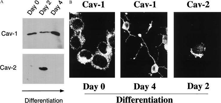
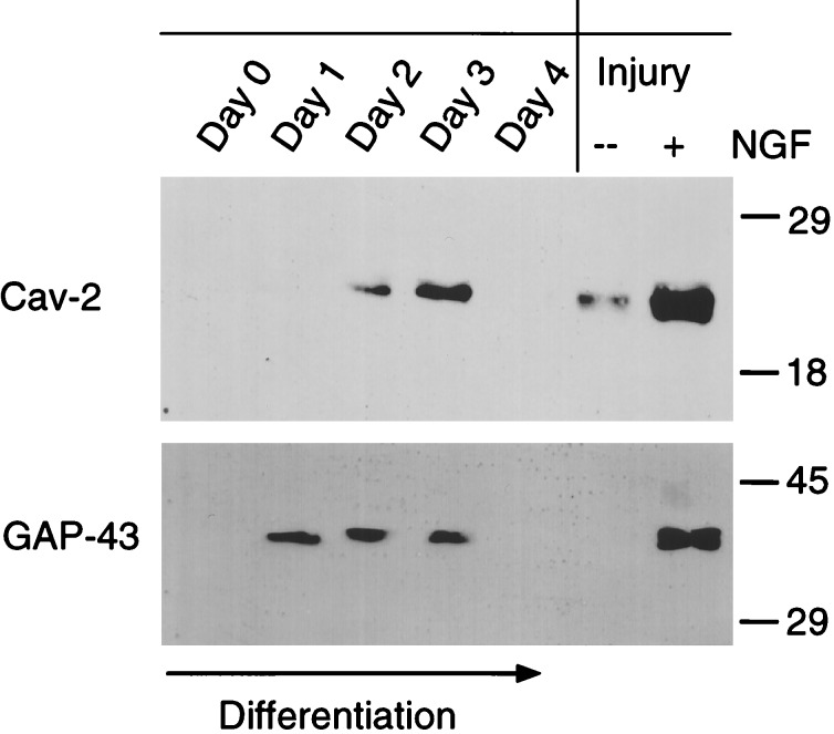
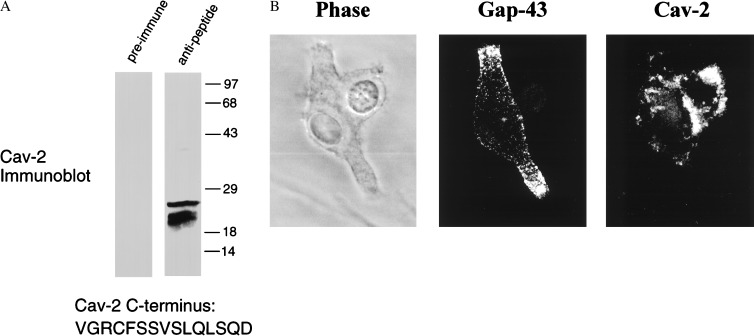
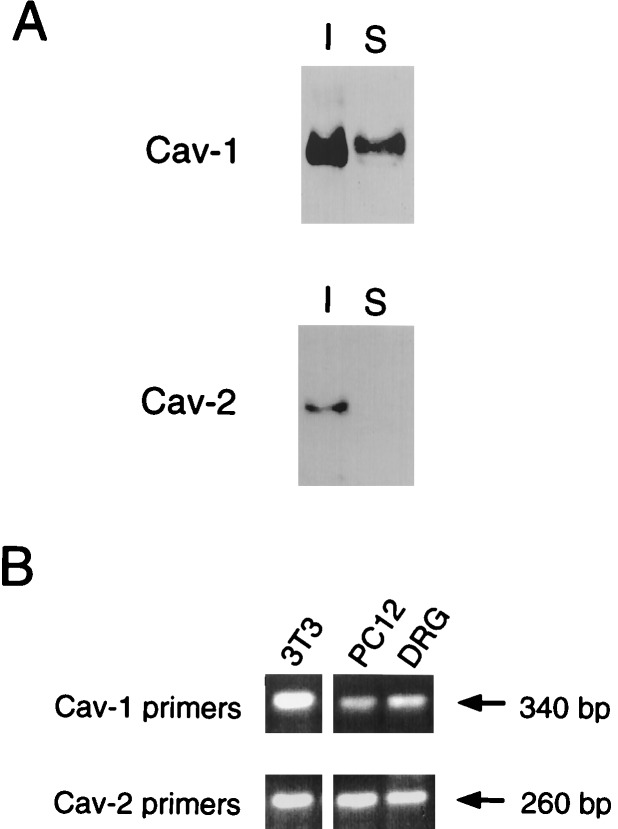
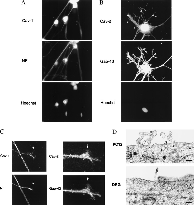
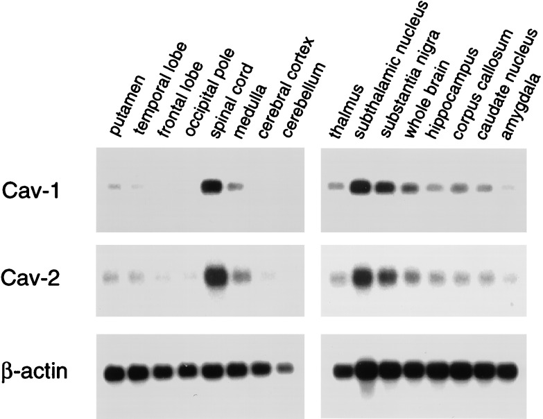
Caveolae are cholesterol/sphingolipid-rich microdomains of the plasma membrane that have been implicated in signal transduction and vesicular trafficking. Caveolins are a family of caveolae-associated integral membrane proteins. Caveolin-1 and -2 show the widest range of expression, whereas caveolin-3 expression is restricted to muscle cell types. It has been previously reported that little or no caveolin mRNA species are detectable in the brain by Northern blot analyses or in neuroblastoma cell lines. However, it remains unknown whether caveolins are expressed within neuronal cells. Here we demonstrate the expression of caveolin-1 and -2 in differentiating PC12 cells and dorsal root ganglion (DRG) neurons by using mono-specific antibody probes. In PC12 cells, caveolin-1 expression is up-regulated on day 4 of nerve growth factor (NGF) treatment, whereas caveolin-2 expression is transiently up-regulated early in the differentiation program and then rapidly down-regulated. Interestingly, caveolin-2 is up-regulated in response to the mechanical injury of differentiated PC12 cells; up-regulation of caveolin-2 under these conditions is strictly dependent on continued treatment with NGF. Robust expression of caveolin-1 and -2 is also observed along the entire cell surface of DRG neurons, including high levels on growth cones. These findings demonstrate that neuronal cells express caveolins.


Figures

Figure 1
Figure 1
Regulated expression of caveolins 1 and 2 during NGF-induced differentiation of PC12 cells. (A) Western Blot analysis. Lysates were prepared from undifferentiated (day 0) and differentiating (days 2 and 4) PC12 cells and subjected to immunoblot analysis with caveolin-1 (pAb N-20) and caveolin-2 (mAb 65) specific antibody probes. Note that caveolin-1 is induced ≈2-fold between days 2 and 4. In contrast, caveolin-2 is transiently up-regulated on day 2 and down-regulated again by day 4. Equal amounts of protein were loaded in each lane. (B) Immunolocalization. Caveolin-1 (pAb N-20) and caveolin-2 (mAb 65) specific antibody probes were used to localize caveolin-1 on days 0 and 4 (Left and Center) and caveolin-2 on day 2 (Right) of differentiation. Note that both caveolins 1 and 2 are most highly expressed on the soma or cell body and less apparent within cellular projections.
Figure 2
Figure 2
Both caveolin-2 and GAP-43 are transiently up-regulated during PC12 cell differentiation; effects of cellular injury. (Left) Lysates were prepared from undifferentiated (day 0) and differentiating (days 1, 2, 3, and 4) PC12 cells and subjected to immunoblot analysis with anti-caveolin-2 (mAb 65) and GAP-43 (mAb; clone GAP-7B10) specific mAb probes. Note that both caveolin-2 and GAP-43 are transiently up-regulated between days 1 and 3 of differentiation and then both are down-regulated on day 4. (Right) After 4 days of differentiation, PC12 cells were trypsinized, subjected to physical injury by repeating pipeting, and replated in the absence (−) or presence (+) of NGF and allowed to recover for 2 days. Lysates were then prepared and subjected to immunoblot analysis with anti-caveolin-2 (mAb 65) and GAP-43 (mAb; clone GAP-7B10). Note that both caveolin-2 and GAP-43 are up-regulated in response to cellular injury and recovery in NGF. Equal amounts of protein were loaded in each lane.
Figure 3
Figure 3
Dual immunolabeling of GAP-43 and caveolin-2 in differentiating PC12 cells. (A) Characterization of an antipeptide antibody directed against the extreme C terminus of caveolin-2. An antipeptide antibody was raised against the peptide VGRCFSSVSLQLSQD that is derived from the extreme C terminus of human caveolin-2 (pAb C-15). Pre-immune and immune serum were incubated in the presence of protein A-Sepharose to purify the IgGs and used to immunoprecipitate a muscle cell lysate that is known to contain caveolin-2. Immunoprecipitates were subjected to Western blot analysis with an anti-caveolin-2 (mAb 65) mAb probe. Note that this caveolin-2 antipeptide antibody (pAb C-15) can be used to immunoprecipitate caveolin-2 from a cellular extract. (B) Dual immunolabeling of GAP-43 and caveolin-2. Caveolin-2 (pAb C-15) and GAP-43 (mAb; clone GAP-7B10) specific antibody probes were used to localize these proteins on day 3 of differentiation. Note that caveolin-2 and GAP-43 do not colocalize. GAP-43 is most highly expressed within the developing cellular projections, whereas caveolin-2 is localized more closely to the soma or cell body and absent from the projections. (Left) Phase image. (Center) GAP-43 immunostaining. (Right) Caveolin-2 immunostaining.
Figure 4
Figure 4
Expression of caveolins 1 and 2 in DRG neurons. (A) Immunoblot analysis. Lysates were prepared from cultures of rat DRG neurons and divided into Triton-soluble (S) and Triton-insoluble (I) fractions. Caveolin-1 (pAb N-20) and caveolin-2 (mAb 65) specific antibody probes were then used to probe these cellular extracts by immunoblotting. Note that both caveolins 1 and 2 are expressed and behave as Triton-insoluble components. (B) RT-PCR analysis. Total RNA isolated from PC12 cells and DRG neurons was subjected to RT-PCR analysis by using primers directed against the known sequences of caveolins 1 and 2; NIH 3T3 cells served as a postitive control.
Figure 5
Figure 5
Localization of caveolins 1 and 2 in cultured DRG neurons. (A and B) Caveolin-1 [pAb N-20 (A)] and caveolin-2 [mAb 65 (B)] specific antibody probes were used to localize caveolins 1 and 2 in cultures of DRG neurons. Neurons were counterstained with antineurofilament protein (NF) or anti-GAP-43 antibodies to identify neuronal cells. Nuclei were visualized by Hoechst staining. (C). Expression of caveolins 1 and 2 in the growth cones of DRG neurons. Arrowheads point at growth cones showing double-labeling with anti-caveolin-1 and antineurofilament antibodies; and double-labeling with anti-caveolin-2 and GAP-43 antibodies. (D). Caveolae-like structures in PC12 cells and DRG neurons. Caveolae were identified by their size (50–100 nm in diameter) and location at or near the plasma membrane. (Bar = 200 nm.)
Figure 6
Figure 6
Distribution of caveolins 1 and 2 in the brain. mRNA species corresponding to caveolins 1 and 2 showed a similar pattern of expression and were highest in the spinal cord, subthalamic nucleus, and substantia nigra, relative to whole brain. The distribution of β-actin mRNA is shown as a control for equal loading.

Similar articles

Cited by

References

    1. Lisanti M P, Scherer P, Tang Z-L, Sargiacomo M. Trends Cell Biol. 1994;4:231–235. - PubMed
    1. Couet J, Li S, Okamoto T, Scherer P S, Lisanti M P. Trends Cardiovasc Med. 1997;7:103–110. - PubMed
    1. Okamoto T, Schlegel A, Scherer P E, Lisanti M P. J Biol Chem. 1998;273:5419–5422. - PubMed
    1. Scherer P E, Lewis R Y, Volonté D, Engelman J A, Galbiati F, Couet J, Kohtz D S, van Donselaar E, Peters P, Lisanti M P. J Biol Chem. 1997;272:29337–29346. - PubMed
    1. Song K S, Scherer P E, Tang Z-L, Okamoto T, Li S, Chafel M, Chu C, Kohtz D S, Lisanti M P. J Biol Chem. 1996;271:15160–15165. - PubMed

Publication types

MeSH terms

LinkOut - more resources