

doi: 10.1073/pnas.95.17.10283.
Bursting calcium rotors in cultured cardiac myocyte monolayers
Affiliations
- PMID: 9707639
- PMCID: PMC21500
- DOI: 10.1073/pnas.95.17.10283
Item in Clipboard
Bursting calcium rotors in cultured cardiac myocyte monolayers
Proc Natl Acad Sci U S A.
.
Abstract
Rotating waves (rotors) of cellular activity were observed in nonconfluent cultures of embryonic chick heart cells by using a macroscopic imaging system that detected fluorescence from intracellular Ca2+. Unlike previous observations of rotors or spiral waves in other systems, the rotors did not persist but exhibited a repetitive pattern of spontaneous onset and offset leading to a bursting rhythm. Similar dynamics were observed in a cellular automaton model of excitable media that incorporates spontaneous initiation of activity, and a decrease of excitability as a consequence of rapid activity (fatigue). These results provide a mechanism for bursting dynamics in normal and pathological biological processes.
Figures
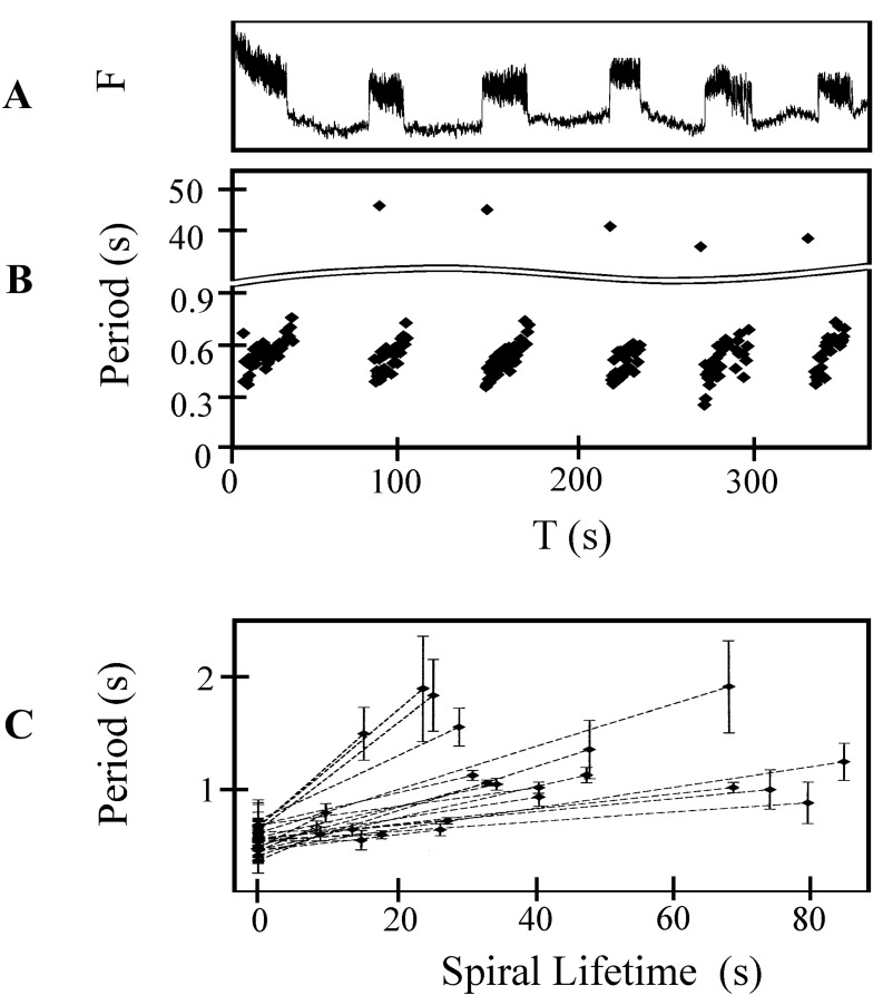
Dynamics of burst generation. (A) Calcium fluorescence for six consecutive bursts from a 1 mm2 area. (B) The interbeat period measured from calcium fluorescence during rotor bursting as a function of time. During each burst duration increases from about 0.4 s to 0.8 s. The interburst duration ranges from 36 s to 44 s. (C) Average period of rotor rotation for 24 representative rotor bursts taken from 6 different preparations. The average period, calculated for the first and last five rotations of each rotor during a burst, is plotted as a function of burst duration. The error bars show the standard deviation of the period over the five averaged periods.
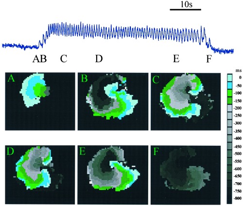
Anatomy of a typical burst. The upper trace shows fluorescence intensity during a single burst. The width of each panel is 1 cm. The colored images show contour plots of activation times at several different times during the burst indicated by the labels. Contour maps are constructed by determining the location of the activation front at 50-ms intervals. The location of the activation front at a given time is plotted in a color given by the key. This format allows for the representation of a number of consecutive raw data frames onto a single composite image. Activation front detection is determined by a threshold set at half maximum fluoresence. (A) Unidirectional block. The wave is initiated at a single site and propagates only to the left. (B) Formation of two mirror-image rotor waves. The excitation doubles back, to invade the tissue forming a mirror-image pair or rotors. (C and D) Contour plots of the mirror-image pair of rotors. (E) Destruction of one of the rotors leaves a single rotor (F) Termination of the remaining rotor.
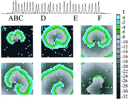
Simulation of a single burst in a 30 × 30 array. Contour maps of activation times for the cellular automaton model during a rotor burst. (A) Unidirectional block. The wave is initiated from the random firing of more than two neighboring sites. Heterogeneity results in unidirectional propagation upward. (B and C) Formation of two mirror-image rotors. The excitation doubles back and invades the site of block to form a rotor. (D and E) Rotation of the mirror-image pair of rotors. The rightmost rotor dominates and the leftmost rotor dies out first. (F) Termination of the remaining rotor.
References
-
- Frazier W T, Kandel E C, Kupfermann I, Waziri R, Coggeshall R E. J Neurophysiol. 1967;30:1288–1351. - PubMed
-
- Canavier C C, Clark J W, Byrne J H. J Neurophysiol. 1991;66:2107–2124. - PubMed
-
- Cook D L, Satin L S, Hopkins W F. Trends Neurosci. 1991;14:411–414. - PubMed
-
- Wang X-J, Rinzel J. In: The Handbook of Brain Theory and Neural Networks. Arbib M A, editor. Cambridge, MA: MIT Press; 1995. pp. 686–691.
Publication types
MeSH terms
Substances
LinkOut - more resources
Full Text Sources
Miscellaneous
