

Differential expression of the peroxisome proliferator-activated receptor gamma (PPARgamma) and its coactivators steroid receptor coactivator-1 and PPAR-binding protein PBP in the brown fat, urinary bladder, colon, and breast of the mouse
- PMID: 9708794
- PMCID: PMC1852994
- DOI: 10.1016/s0002-9440(10)65577-0
Differential expression of the peroxisome proliferator-activated receptor gamma (PPARgamma) and its coactivators steroid receptor coactivator-1 and PPAR-binding protein PBP in the brown fat, urinary bladder, colon, and breast of the mouse
Abstract
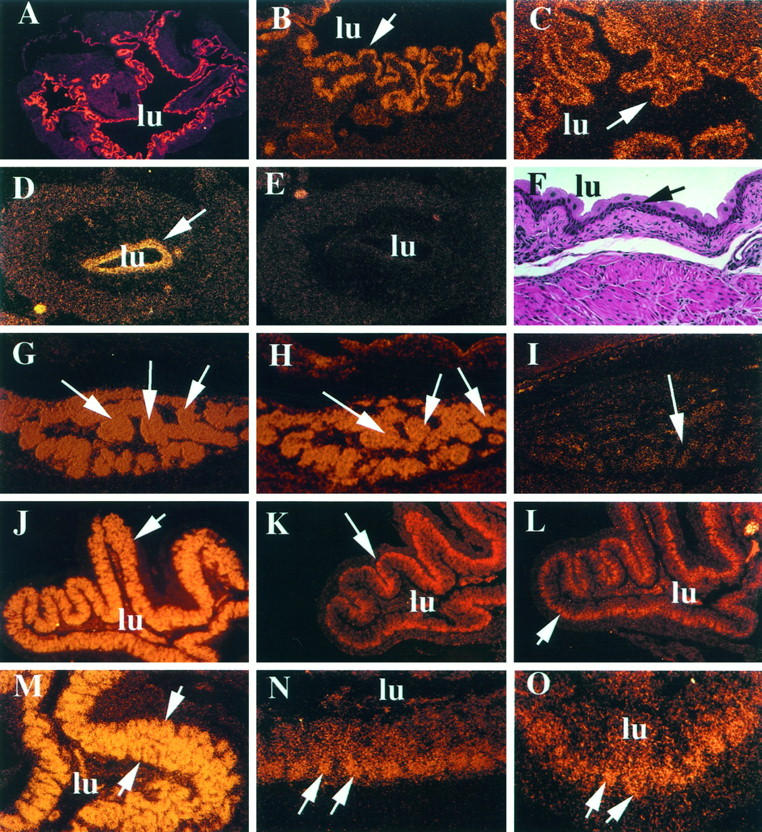
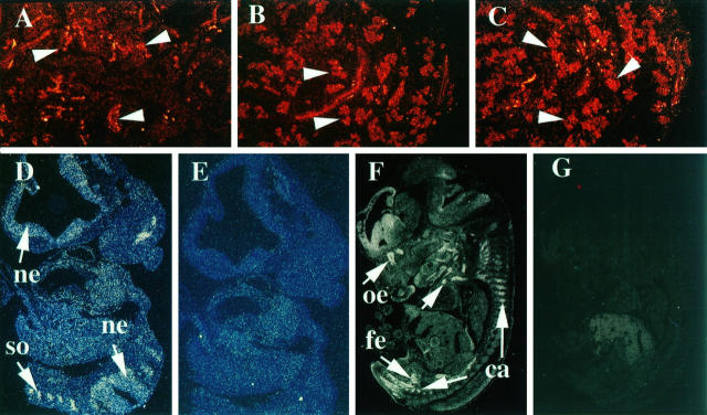
Peroxisome proliferator-activated receptors (PPARs) regulate genes involved in lipid metabolism and adipocyte differentiation. Steroid receptor coactivator-1 (SRC-1) and PPAR-binding protein (PBP) interact with PPARgamma and act as coactivators to enhance ligand-dependent transcription. We report here that PPARgamma, SRC-1, and PBP are differentially expressed in the brown fat, transitional epithelium of the urinary bladder, colonic mucosa, and mammary epithelium of the adult mouse. PPARgamma and PBP are expressed in the transitional epithelium of urinary bladder and in brown adipose tissue, but not SRC-1. In the colonic mucosa, PPARgamma expression occurs throughout the villi, whereas the expression of both SRC-1 and PBP is confined mostly to the crypts. The expression of both SRC-1 and PBP is prominent in the breast epithelium of nonpregnant, pregnant, and lactating mice, whereas PPARgamma expression appeared prominent during lactation. During early embryonic development, PPARgamma, SRC-1, and PBP are differentially expressed, with only limited cell types displaying overlapping expression. PPARgamma and PBP expression overlapped in the brown fat and urogenital sinus at stage E15.5 of embryogenesis, whereas SRC-1 expression occurred mostly in neuroepithelium and cartilage between stages E9.5 and E13.5 of embryogenesis.
Figures
Similar articles
-
Isolation and characterization of PBP, a protein that interacts with peroxisome proliferator-activated receptor.J Biol Chem. 1997 Oct 10;272(41):25500-6. doi: 10.1074/jbc.272.41.25500. J Biol Chem. 1997. PMID: 9325263
-
Discrete roles for peroxisome proliferator-activated receptor gamma and retinoid X receptor in recruiting nuclear receptor coactivators.Mol Cell Biol. 2000 Nov;20(21):8008-17. doi: 10.1128/MCB.20.21.8008-8017.2000. Mol Cell Biol. 2000. PMID: 11027271 Free PMC article.
-
Transcription coactivator PBP, the peroxisome proliferator-activated receptor (PPAR)-binding protein, is required for PPARalpha-regulated gene expression in liver.J Biol Chem. 2004 Jun 4;279(23):24427-34. doi: 10.1074/jbc.M402391200. Epub 2004 Mar 29. J Biol Chem. 2004. PMID: 15150259
-
Peroxisome proliferator-activated receptors, coactivators, and downstream targets.Cell Biochem Biophys. 2000;32 Spring:187-204. doi: 10.1385/cbb:32:1-3:187. Cell Biochem Biophys. 2000. PMID: 11330046 Review.
-
Development, validation and implementation of an in vitro model for the study of metabolic and immune function in normal and inflamed human colonic epithelium.Dan Med J. 2015 Jan;62(1):B4973. Dan Med J. 2015. PMID: 25557335 Review.
Cited by
-
Coactivators in PPAR-Regulated Gene Expression.PPAR Res. 2010;2010:250126. doi: 10.1155/2010/250126. Epub 2010 Aug 5. PPAR Res. 2010. PMID: 20814439 Free PMC article.
-
Expression and role of nuclear receptor coregulators in colorectal cancer.World J Gastroenterol. 2017 Jul 7;23(25):4480-4490. doi: 10.3748/wjg.v23.i25.4480. World J Gastroenterol. 2017. PMID: 28740336 Free PMC article. Review.
-
Activation of peroxisome proliferator-activated receptor-gamma reverses squamous metaplasia and induces transitional differentiation in normal human urothelial cells.Am J Pathol. 2004 May;164(5):1789-98. doi: 10.1016/s0002-9440(10)63737-6. Am J Pathol. 2004. PMID: 15111325 Free PMC article.
-
Peroxisome proliferator-activated receptor gamma controls Muc1 transcription in trophoblasts.Mol Cell Biol. 2004 Dec;24(24):10661-9. doi: 10.1128/MCB.24.24.10661-10669.2004. Mol Cell Biol. 2004. PMID: 15572671 Free PMC article.
-
Peroxisome Proliferator-Activated Receptor-gamma in Amyotrophic Lateral Sclerosis and Huntington's Disease.PPAR Res. 2008;2008:418765. doi: 10.1155/2008/418765. PPAR Res. 2008. PMID: 18464922 Free PMC article.
References
-
- Tolbert NE: Metabolic pathways in peroxisomes and glyoxisomes. Annu Rev Biochem 1981, 50:133-157 - PubMed
-
- Reddy JK, Krishnakantha TP: Hepatic peroxisome proliferation: induction by two novel compounds structurally unrelated to clofibrate. Science 1975, 200:787-789 - PubMed
-
- Reddy JK, Lalwani ND: Carcinogenesis by hepatic peroxisome proliferators: evaluation of the risk of hypolipidemic drugs and plasticizers to human. CRC Crit Rev Toxicol 1983, 12:1-58 - PubMed
-
- Reddy JK, Goel SK, Nemali MR, Carrino JJ, Laffler TG, Reddy MK, Sperbeck SJ, Osumi T, Hashimoto T, Lalwani ND, Rao MS: Transcriptional regulation of peroxisomal fatty acyl-CoA oxidase and enoyl-CoA hydratase/3-hydroxyacyl-CoA dehydrogenase in rat liver by peroxisome proliferators. Proc Natl Acad Sci USA 1986, 83:1747-1751 - PMC - PubMed
-
- Issemann I, Green S: Activation of a member of the steroid hormone receptor superfamily by peroxisome proliferators. Nature 1990, 347:645-650 - PubMed
Publication types
MeSH terms
Substances
Grants and funding
LinkOut - more resources
Full Text Sources
Molecular Biology Databases
Research Materials
Miscellaneous
