. 1998 Aug;153(2):395-403.
doi: 10.1016/S0002-9440(10)65583-6.

Expression of the vascular endothelial growth factor C receptor VEGFR-3 in lymphatic endothelium of the skin and in vascular tumors

Affiliations

Expression of the vascular endothelial growth factor C receptor VEGFR-3 in lymphatic endothelium of the skin and in vascular tumors

A Lymboussaki et al. Am J Pathol. 1998 Aug.

Abstract

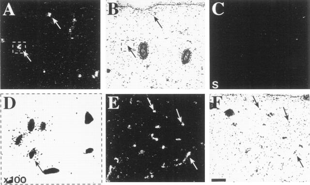
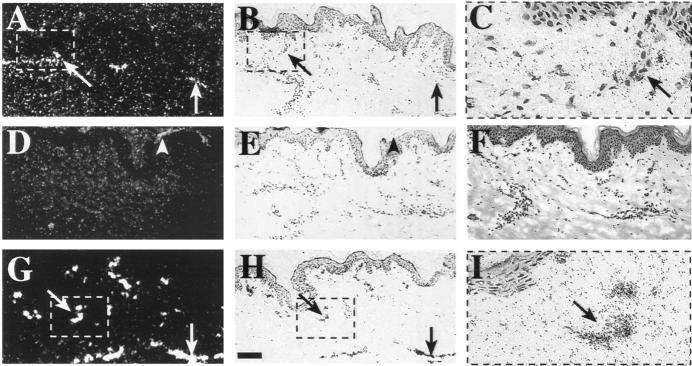
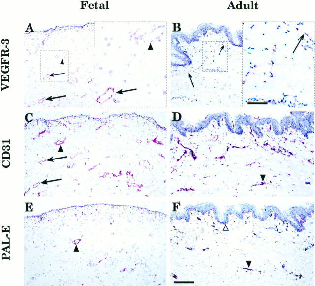
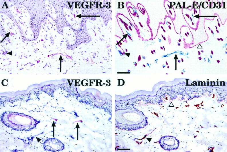
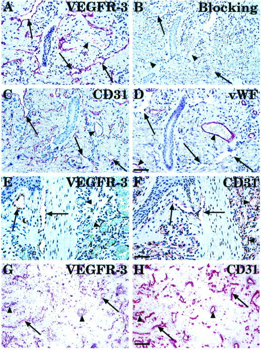
It is difficult to identify lymph vessels in tissue sections by histochemical staining, and thus a specific marker for lymphatic endothelial cells would be more practical in histopathological diagnostics. Here we have applied a specific antigenic marker for lymphatic endothelial cells in the human skin, the vascular endothelial growth factor receptor-3 (VEGFR-3), and show that it identifies a distinct vessel population both in fetal and adult skin, which has properties of lymphatic vessels. The expression of VEGFR-3 was studied in normal human skin by in situ hybridization, iodinated ligand binding, and immunohistochemistry. A subset of developing vessels expressed the VEGFR-3 mRNA in fetal skin as shown by in situ hybridization and radioiodinated vascular endothelial growth factor (VEGF)-C bound selectively to a subset of vessels in adult skin that had morphological characteristics of lymphatic vessels. Monoclonal antibodies against the extracellular domain of VEGFR-3 stained specifically endothelial cells of dermal lymph vessels, in contrast to PAL-E antibodies, which stained only blood vessel endothelia. In addition, staining for VEGFR-3 was strongly positive in the endothelium of cutaneous lymphangiomatosis, but staining of endothelial cells in cutaneous hemangiomas was weaker. These results establish the utility of anti-VEGFR-3 antibodies in the identification of lymphovascular channels in the skin and in the differential diagnosis of skin lesions involving lymphatic or blood vascular endothelium.


Figures

Figure 1.
Figure 1.
Expression of VEGFR-3 and VEGFR-2 in fetal skin analyzed by in situ hybridization. A, B, and D: Hybridization with the VEGFR-3 antisense probe. The signal originates from a subset of vessels of the dermis (arrows). C: Control hybridization with the VEGFR-3 sense strand. D: In a higher magnification of the area marked in (A) and (B), the autoradiographic grains are shown to be localized to the endothelial cells of a developing vessel. E and F: Hybridization for VEGFR-2. The probe gives signal in developing vessel structures at several levels of the dermis (arrows). B, D, and F: Bright-field photographs of the same sections. Bar: 0.1 mm.
Figure 2.
Figure 2.
Radioactive VEGF-C and VEGF binding in human adult skin visualized by autoradiography. A, B, and C: Binding of 125I-labeled rh-VEGF-C. The signal is localized to the thin lymphatic vessels of the subpapillary plexus (arrows). D, E, and F: Binding sites are blocked when a 100-fold excess of cold ligand is added to the incubation medium. Some unspecific binding is seen originating from keratin squames in the epidermis (D, arrowhead). G, H, and I: Specific 125I-labeled rh-VEGF binding to vascular endothelium, which extends throughout the dermis. (C and I: Higher magnifications (×100) of the marked vessel structures binding the radioactive ligands VEGF-C and VEGF, respectively. Dark-field (A, D, and G) and bright-field (B, C, E, F, H, and I) exposures are shown. Bar: 0.1 mm for A, B, and D to H.
Figure 3.
Figure 3.
Comparison of VEGFR-3 expression with two vascular endothelial markers in fetal and adult skin. Arrows indicate vessels in adjacent sections of fetal and adult skin, respectively, which stain for VEGFR-3 (A and B) and CD31 (C and D), but not for PAL-E (E and F), and are therefore presumably lymphatic. Arrowheads identify vessels, which only react with antibodies against the two blood vascular endothelial antigens. In adult skin, the thin VEGFR-3-positive lymphatic vessels could be identified by morphology and lack of a basal lamina (B), but the same vessel could not be traced in the adjacent sections stained for CD31 (D) or PAL-E (F). Bar: 0.17 mm. Insets (A and B, right) illustrate higher magnifications of the lymphatic vessels (arrows) in marked areas of the fetal and adult skin sections, respectively. Bar: 57 μm.
Figure 4.
Figure 4.
Comparison of VEGFR-3 expression with double staining for two vascular endothelial markers and with anti-laminin staining. A sample of adult buccal mucosa was stained with the anti-VEGFR-3 mAb (A) and an adjacent section, first with PAL-E (B, red-colored endothelia) and then with anti-CD31 (B, blue-colored endothelia), to distinguish lymphatic structures from blood vessels. PAL-E also decorates the epidermal basal lamina (B, open triangle). Comparison of VEGFR-3 and laminin expression in adult skin is shown in (C) and (D). Arrows indicate lymphatic vessels of adult skin, which stain for VEGFR-3 (C). Arrowheads show blood vessels with basement membranes, which stain for laminin (D), but only very weakly for VEGFR-3. Bars: 35 μm (A and B) and 0.11 mm (C and D).
Figure 5.
Figure 5.
Comparison of VEGFR-3 expression with vascular endothelial markers in lymphangiomatosis (A to D) and in capillary cutaneous (E and F) and intramuscular (G and H) hemangiomas. Arrows indicate collapsed, VEGFR-3-positive thin endothelium-lined anastomosing channels dissecting through dermal connective tissue in a lymphangiomatosis sample (A). Negative control was done by blocking anti-VEGFR-3 with the immunogen (B). Adjacent sections were also stained for CD31 (C) and vWF (D). Note that in contrast to the buccal mucosa sample (Figure 4) ▶ , CD31 identifies the endothelial cells lining lymphatic spaces in lymphangiomatosis, whereas the vWF staining is weak or absent. In cutaneous hemangioma, the well-formed capillaries with prominent endothelial cells show very little or no expression of VEGFR-3 (E), but are strongly CD31 positive (F). In a frozen section of intramuscular hemangioma, weak VEGFR-3 staining is detected (G, arrows) in part of the CD31-positive vessels (H). Bars: 65 μm (A to D, 48 μm (E and F), and 140 μm (G and H).

Similar articles

Cited by

References

    1. Folkman J, Shing Y: Angiogenesis. J Biol Chem 1992, 267:10931-10934 - PubMed
    1. Hanahan D, Folkman J: Patterns and emerging mechanisms of the angiogenic switch during tumorigenesis. Cell 1996, 86:353-364 - PubMed
    1. Ferrara N, Davis-Smyth T: The biology of vascular endothelial growth factor. Endocr Rev 1997, 18:4-25 - PubMed
    1. Maglione D, Guerriero V, Viglietto G, Delli-Bovi P, Persico MG: Isolation of a human placenta cDNA coding for a protein related to the vascular permeability factor. Proc Natl Acad Sci USA 1991, 88:9267-9271 - PMC - PubMed
    1. Joukov V, Pajusola K, Kaipainen A, Chilov D, Lahtinen I, Kukk E, Saksela O, Kalkkinen N, Alitalo K: A novel vascular endothelial growth factor, VEGF-C, is a ligand for the Flt4 (VEGFR-3) and KDR (VEGFR-2) receptor tyrosine kinases. EMBO J 1996, 15:290-298 - PMC - PubMed

Publication types

MeSH terms

Substances