. 1998 Aug;153(2):417-28.
doi: 10.1016/S0002-9440(10)65585-X.

Coxsackievirus B3-induced myocarditis: perforin exacerbates disease, but plays no detectable role in virus clearance

Affiliations

Coxsackievirus B3-induced myocarditis: perforin exacerbates disease, but plays no detectable role in virus clearance

J R Gebhard et al. Am J Pathol. 1998 Aug.

Abstract

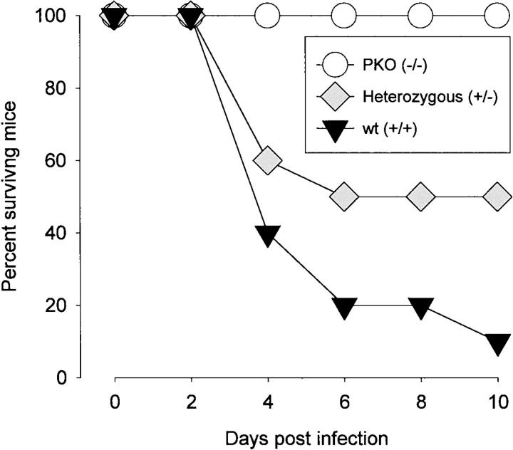
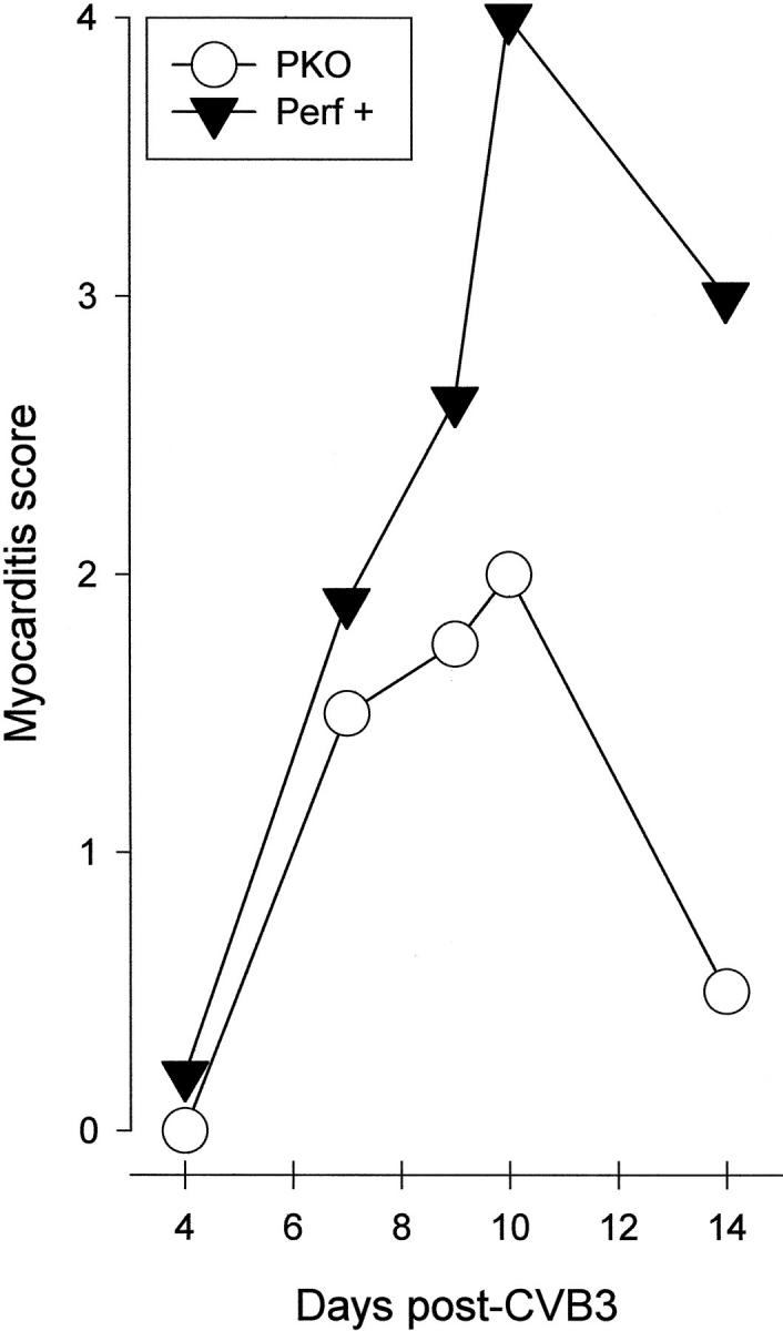
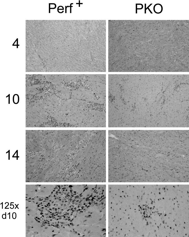
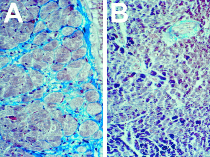
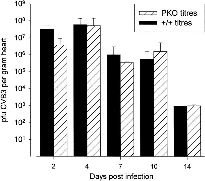
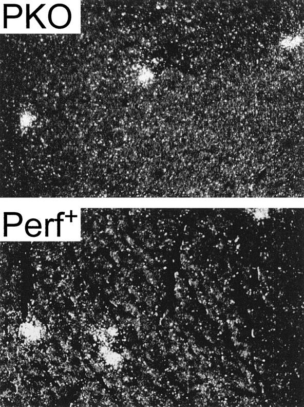
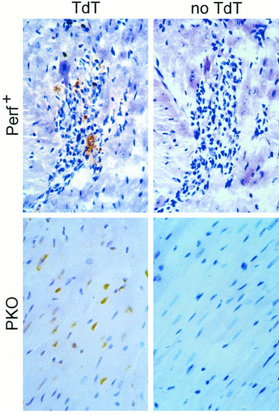
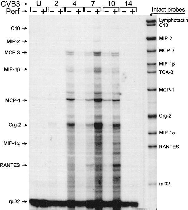
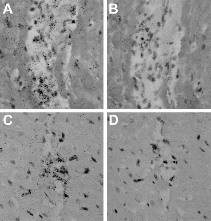
Viral myocarditis is remarkably common, being detected in approximately 1% of unselected asymptomatic individuals. Many cases are attributable to enteroviral infection, and in particular to coxsackievirus B3. The underlying pathogenesis is controversial, but most studies admit the important immunopathological role of infiltrating CD8+ (cytotoxic) T lymphocytes (CTLs). We have previously shown that CTLs play conflicting roles in coxsackievirus B (CVB) myocarditis; they assist in controlling virus replication, but also are instrumental in causing the extensive inflammatory disease, which often results in severe myocardial scarring. A role for perforin, the major CTL cytolytic protein, in CVB myocarditis has been suggested, but never proven. In the present study we use perforin knockout (PKO) mice to show that perforin plays a major role in CVB infection; in broad terms, perforin is important in immunopathology, but not in CVB clearance. For example, PKO mice are better able to withstand a normally lethal dose of CVB (100% survival of PKO mice compared with 90% death in +/+ littermates). In addition, PKO mice given a nonlethal dose of CVB develop only a mild myocarditis, whereas their perforin+ littermates have extensive myocardial lesions. The myocarditis in PKO mice resolves more quickly, and these mice show minimal histological sequelae; in contrast, late in disease the perforin+ mice develop severe myocardial fibrosis. PKO mice, despite lacking this major CTL effector function, can control the infection and eradicate the virus; growth kinetics and peak CVB titers are indistinguishable in PKO and perforin+ mice. Therefore, the immunopathological and antiviral effects of CTLs can be uncoupled by ablation of perforin; this offers a promising target for therapy of myocarditis. Furthermore, we evaluate the possible roles of apoptosis, and of chemokine expression, in CVB infection. In perforin+ mice, apoptotic cells are detected within the inflammatory infiltrate, whereas in their PKO counterparts, apoptotic myocyte nuclei are seen. Chemokine expression in both PKO and perforin+ mice precedes and parallels the course of myocarditis. Several chemokines are detectable earlier in PKO mice than in perforin+ mice, but PKO mice show reduced peak levels, and chemokine expression decays sooner. In particular, MIP-1alpha expression is barely detectable at any time point in PKO mice, but it is readily identified in perforin+ animals, peaking just before the time of maximal myocarditis; this is particularly interesting, given that MIP-1alpha knockout mice are resistant to CVB myocarditis, but remain able to control viral infection. Thus, the chemokine pathway offers a second route of intervention to diminish myocarditis and its sequelae, while permitting the host to eradicate the virus.


Figures

Figure 1.
Figure 1.
Perforin contributes to lethal outcome of CVB3 infection. Three groups of male mice (10 mice per group) were infected with CVB3 (100 pfu intraperitoneally) and were observed daily for 21 days. No deaths occurred after day 10 p.i.
Figure 2.
Figure 2.
Myocarditis index in PKO and perforin+ mice. Mice were infected with 35 pfu of CVB3 and were sacrificed at the indicated time points (Days post-CVB3). Hearts were harvested, fixed, paraffin embedded and sectioned, and stained with H&E. Inflammatory lesions were counted. The scoring criteria are described in Materials and Methods and reflect only the number of lesions, rather than the total extent of myocardial involvement.
Figure 3.
Figure 3.
Myocarditis in perforin+ and PKO mice: perforin is required for extensive infiltration. Representative H&E-stained sections of hearts from CVB-infected perforin+ mice (left) and PKO mice (right) at days 4, 10, and 14 p.i. Original magnification, ×50. The final row shows enlargements (original magnification, ×25) of myocardial lesions at day 10 p.i.
Figure 4.
Figure 4.
Reduced fibrosis in hearts of PKO mice. Representative sections from hearts of perforin+ mice (A) and PKO littermates (B) taken 30 to 40 days p.i., at which time no detectable infectious virus is present. In all cases, 35 pfu CVB was administered, and virus infection and replication were confirmed by serum titration during the first 10 days p.i. Sections are stained with Masson’s trichrome, which stains normal cardiac muscle cells red/purple and stains fibrous tissue light blue.
Figure 5.
Figure 5.
Perforin has no detectable effect on CVB3 titers or growth kinetics. PKO mice and perforin+ littermates were challenged with CVB3, and heart titers (pfu/g of tissue) were determined at the indicated times. For each strain, each time point represents the average titer of at least four mice; error bars are shown.
Figure 6.
Figure 6.
Similar virus distribution 4 days post-CVB3 infection in PKO and +/− mice. PKO or perforin+ mice were infected with CVB3, and at 4 days p.i. (the peak of virus titer in the heart), mice were sacrificed. Paraffin sections of the myocardia were analyzed by in situ hybridization using an antisense probe for CVB3. After emulsification and development, slides were visualized by dark-field microscopy. Original magnification, ×80.
Figure 7.
Figure 7.
Apoptosis in CVB3-infected myocardium. The hearts harvested at 10 days p.i. and evaluated for myocarditis (shown in Figure 3 ▶ ) were tested for apoptosis using the terminal deoxynucleotidyl transferase (TdT)-mediated nick-end labeling assay. Reactions were carried out in the presence of the enzyme TdT (left; TdT) or, as a control, in its absence (right; no TdT). Signal in perforin+ mice was seen only within the inflammatory foci (top left), whereas in PKO mice signal was absent from the small foci, but was detected in myocyte nuclei (bottom left).
Figure 8.
Figure 8.
Chemokine mRNA levels in hearts of CVB3-infected PKO and perforin+ mice. RNAs were prepared from the hearts of perforin+ and PKO mice at the indicated time points p.i., and from uninfected mice (U). Chemokine mRNAs were detected by RNase protection, as described in Materials and Methods. Intact probes are shown in the right lane, and the positions of the protected bands are indicated (arrows) to the left of the figure, rp132 is a control probe, which detects an mRNA encoding a 32-kb ribosomal protein; this is included to confirm the assay execution and to ensure that all RNA samples were qualitatively and quantitatively comparable.
Figure 9.
Figure 9.
In situ hybridization for chemokine expression. Paraffin sections of hearts from CVB3-infected perforin+ mice (A and B) or PKO mice (C and D) were prepared 7 days p.i. and were analyzed by in situ hybridization using an antisense probe for crg-2 (A and C). As a control, adjacent sections of each heart were hybridized with a sense probe for crg-2 (B and D). After emulsification and development, slides were stained with H&E and were visualized by bright-field microscopy. Original magnification, ×160.

Similar articles

Cited by

References

    1. O’Connell JB: The role of myocarditis in end-stage dilated cardiomyopathy. Tex Heart Inst J 1987, 14:268-275 - PMC - PubMed
    1. Gravanis MB, Sternby NH: Incidence of myocarditis: a 10-year autopsy study from Malmo, Sweden. Arch Pathol Lab Med 1991, 115:390-392 - PubMed
    1. Sole MJ, Liu P: Viral myocarditis: a paradigm for understanding the pathogenesis and treatment of dilated cardiomyopathy. J Am Coll Cardiol 1993, 22:99A-105A - PubMed
    1. Goodwin JF: Cardiomyopathies and specific heart muscle diseases: definitions, terminology, classifications and new and old approaches. Postgrad Med J 1992, 68(suppl 1):S3-S6 - PubMed
    1. Heck CF, Shumway SJ, Kaye MP: The Registry of the International Society for Heart Transplantation: sixth official report, 1989. J Heart Transplant 1989, 8:271-276 - PubMed

Publication types

MeSH terms