. 1998 Aug;153(2):429-37.
doi: 10.1016/S0002-9440(10)65586-1.

Expression of matrix metalloproteinases and their tissue inhibitors in human brain tumors

Affiliations

Expression of matrix metalloproteinases and their tissue inhibitors in human brain tumors

K Lampert et al. Am J Pathol. 1998 Aug.

Abstract

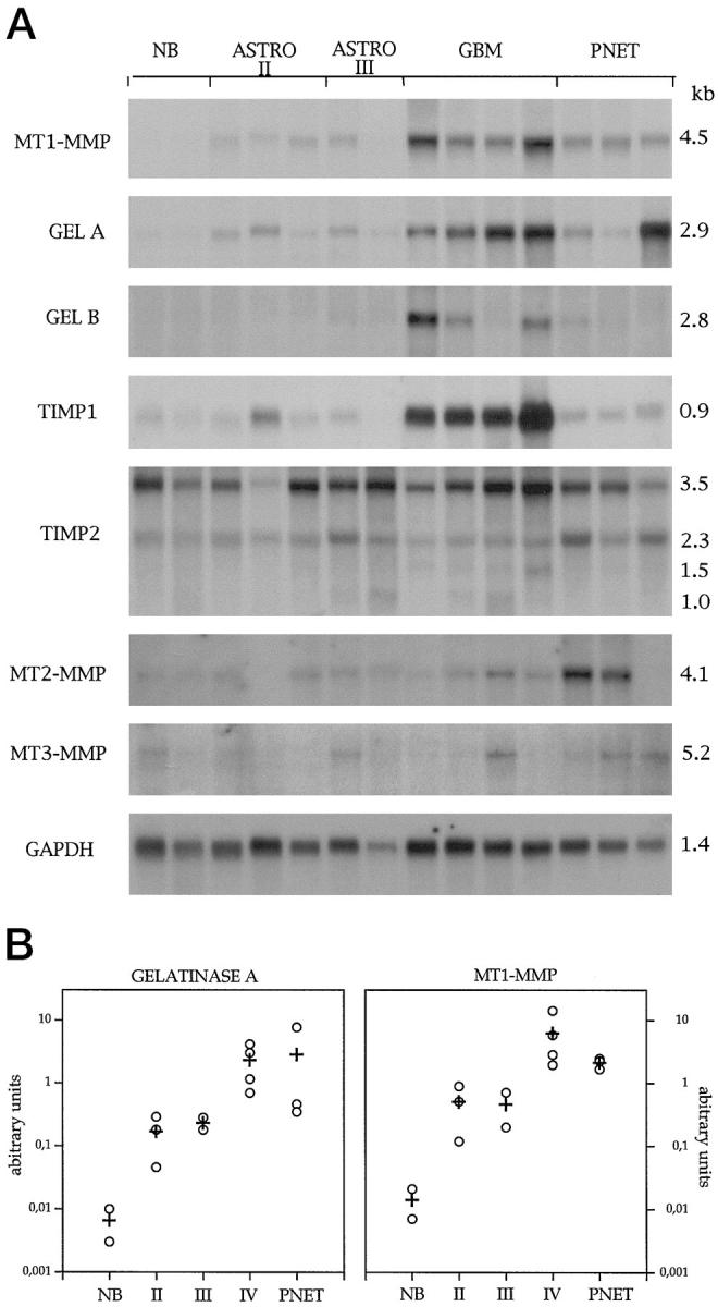
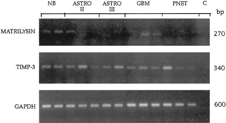
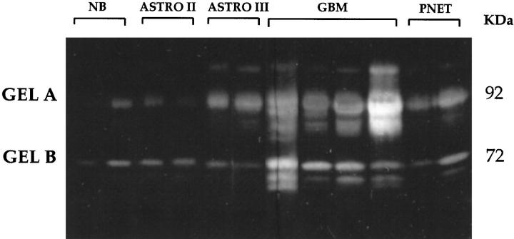
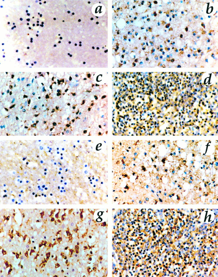
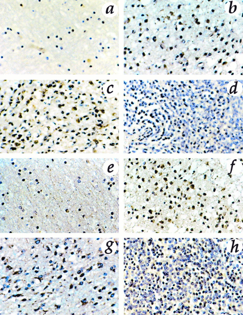
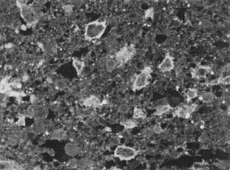
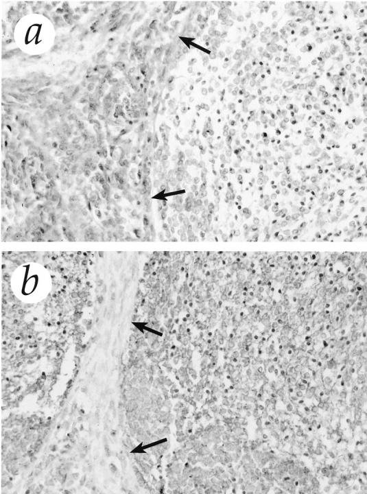
In this study, we investigated the expression patterns of 15 matrix metalloproteinases (MMPs) and three tissue inhibitors of metalloproteinase in gliomas, medulloblastomas, and normal brain tissue. By Northern blot analysis we found increased levels of mRNAs encoding for gelatinase A, gelatinase B, two membrane-type MMPs (mt1- and mt2-MMP), and tissue inhibitors of metalloproteinase-1 in glioblastomas and medulloblastomas. We observed a significant increase of mt1-MMP, gelatinase A, gelatinase B, and tissue inhibitors of metalloproteinase-1 in glioblastomas as compared with low-grade astrocytomas, anaplastic astrocytomas, and normal brain. In medulloblastomas, the expression of mt1-MMP, mt2-MMP, and gelatinase A were also increased, but to a lesser extent than that observed in glioblastomas. These data were confirmed at the protein level by immunostaining analysis. Moreover, substrate gel electrophoresis showed that the activated forms of gelatinases A and B were present in glioblastomas and medulloblastomas. These results suggest that increased expression of mt1-MMP/gelatinase A is closely related to the malignant progression observed in gliomas. Furthermore, the present study demonstrates, to our knowledge for the first time, that medulloblastomas express high levels of MMP.


Figures

Figure 1.
Figure 1.
A:Northern blot analysis of MMP and TIMP mRNAs in normal brain (NB), low-grade astrocytoma (Astro II), anaplastic astrocytoma (Astro III), GBM, and medulloblastoma (PNET). In normal brain, we found high levels of TIMP-2, whereas TIMP-1 and all tested MMPs were expressed at low levels only. In low-grade astrocytomas and anaplastic astrocytoma, an increase in the expression of mt1-MMP, gelatinase A, and TIMP-1 was seen. The expression of these genes increased dramatically in GBM showing concomitant up-regulation of TIMP-1 mRNA. TIMP-2 was expressed in all tissues included in this study. In medulloblastomas, mt1-MMP and gelatinase A were overexpressed. mt2-MMP was up-regulated in two cases of medulloblastoma. B: Relative hybridization numbers (○) and mean (+) of mt1-MMP and gelatinase A mRNAs in normal brain (NB), low-grade astrocytoma (II), anaplastic astrocytoma (III), GBM (IV), and medulloblastoma (PNET). The mRNA levels were quantified by densitometric analysis using an Image Master System DTS (Pharmacia). GAPDH was used as loading control. The relative hybridization signal numbers were calculated by ascribing an arbitrary value of 1 to the least intense signal.
Figure 2.
Figure 2.
PCR analysis of matrilysin and TIMP-3 expression in human brain tumors. Signals appearing in the three lanes of the normal brain were about 350 bp and therefore longer than the expected size of 270 bp. Some weak matrilysin expression was seen in two GBMs. TIMP-3 expression was seen in all tissues. NB, normal brain; Astro II, low-grade astrocytoma; Astro III, anaplastic astrocytoma; PNET, medulloblastoma; C, negative control.
Figure 3.
Figure 3.
Gelatin substrate gel electrophoresis of normal brain, low-grade astrocytomas (Astro II), anaplastic astrocytoma (Astro III), GBM, and medulloblastoma (PNET). Indicated by Mr 92 kd and by Mr 72 kd are the progelatinase B and the progelatinase A, respectively. Constitutive secretion of the latent forms of gelatinases A and B appeared in every tissue examined, including normal brain. In normal brain, low-grade astrocytomas, and anaplastic astrocytoma, no detectable amounts of the activated enzymes were found. In contrast, in GBM, an increase of active forms of gelatinases was clearly demonstrable, as indicated by a second and even a third band. In medulloblastomas, the level of proenzyme forms was high, with only barely detectable active forms of both gelatinases.
Figure 4.
Figure 4.
Immunostaining for gelatinase A (a to d) and mt1-MMP (e to h) in normal brain (a and e), low-grade astrocytoma (b and f), GBM (c and g), and medulloblastoma (d and h). Original magnification, ×313. Normal brain showed no immunoreactivity. Staining was positive for mt1-MMP in tumor cells localized in the cell membrane. Gelatinase A was identified intracytoplasmatically in tumor cells and in partial colocalization with mt1-MMP. In low-grade astrocytomas, few cells displayed a sporadic, positive staining for gelatinase A and mt1-MMP, whereas malignant gliomas and medulloblastomas showed an intense immunoreaction.
Figure 5.
Figure 5.
Immunostaining for gelatinase B (a to d) and TIMP-2 (e to h) in normal brain (a and e), low-grade astrocytoma (b and f), GBM (c and g), and medulloblastoma (d and h). Original magnification, ×313. Positive staining for gelatinase B was localized inside the cytoplasm of tumor cells; it was weak in medulloblastomas and low-grade astrocytomas, whereas glioblastomas were strongly stained. Immunostaining for TIMP-2 showed nests of positive cells in all tumors. Endothelial cells were immunostained with anti-gelatinase B and TIMP-2. TIMP-2 was detected in normal brain primarily in endothelial cells and in few astrocytes and neurons.
Figure 6.
Figure 6.
Immunofluorescence with anti-mt1-MMP antibody and confocal laser microscopy clearly showed immunostaining on the cell membrane of tumor cells. Original magnification, ×400.
Figure 7.
Figure 7.
Immunostaining for gelatinase A (a) and mt1-MMP (b) in desmoplastic medulloblastoma. The high cellular trabeculae of tumor cells (arrows) showed an intense immunostaining for gelatinase A, whereas the reticulin-free islands stained predominantly for mt1-MMP. Original magnification, ×313.

References

    1. Giles GG, Gonzales MF: Epidemiology of brain tumors and factors in prognosis. Kaye AH Laws ER eds. Brain Tumors, ch 4. 1995, :pp 47-67 Churchill Livingstone, New York
    1. Gold EB: Epidemiology of brain tumors. Lilenfeld AM eds. Reviews in Cancer Epidemiology, 1980, vol 1.:pp 245-292 Elsevier, North Holland, the Netherlands
    1. Cerame MA, Guthikonda M, Kohli CM: Extraneural metastases in gliosarcomas: a case report and review of literature. Neurosurgery 1985, 17:413-418 - PubMed
    1. Rutka JT, Dougherty DV, Giblin JR, Edwards MS, McCulloch JR, Rosenblum ML: Growth of a medulloblastoma on normal leptomeningeal cells in culture: interaction of tumor cells and normal cells. Neurosurgery 1987, 21:872-878 - PubMed
    1. Berger MS, Magrassi L, Geyer R: Medulloblastomas and primitive neuroectodermal tumors. Kaye AH Laws ER eds. Brain Tumors, ch 30. 1995, :pp 562-574 Churchill Livingstone, New York

Publication types

MeSH terms