. 1998 Aug;153(2):447-56.
doi: 10.1016/S0002-9440(10)65588-5.

Apoptosis of retrogradely degenerating neurons occurs in association with the accumulation of perikaryal mitochondria and oxidative damage to the nucleus

Affiliations

Apoptosis of retrogradely degenerating neurons occurs in association with the accumulation of perikaryal mitochondria and oxidative damage to the nucleus

N A Al-Abdulla et al. Am J Pathol. 1998 Aug.

Abstract

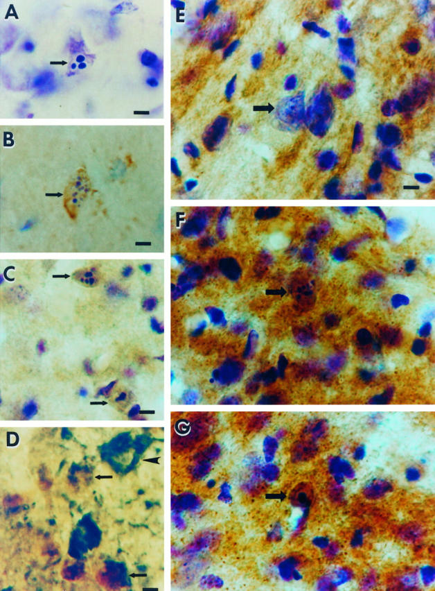
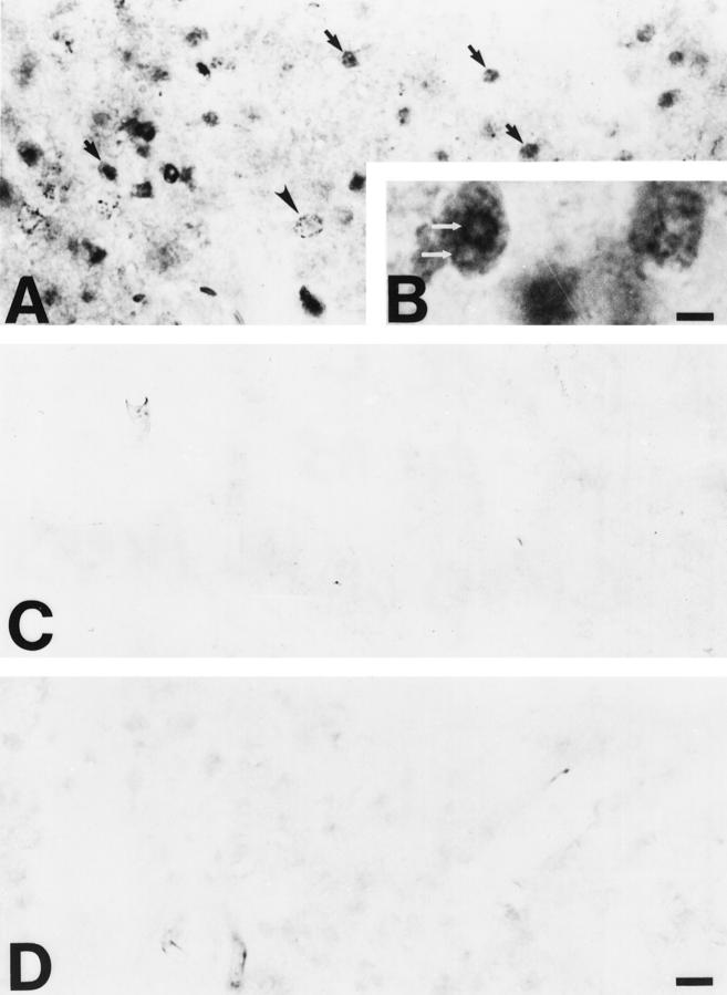
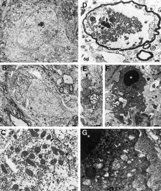
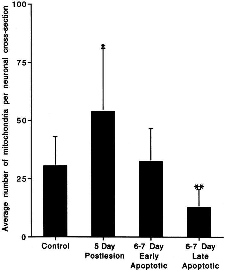
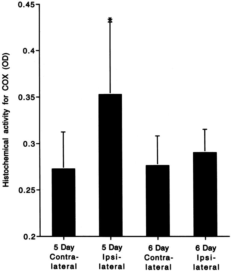
The mechanisms for neuronal apoptosis after axotomy and target deprivation in the adult central nervous system are poorly understood. We used a unilateral occipital cortex ablation model in the adult rat to test the hypothesis that apoptotic retrograde neurodegeneration in the dorsal lateral geniculate nucleus occurs in association with oxidative stress and mitochondrial abnormalities. Immunodetection of 8-hydroxy-2'-deoxyguanosine, a marker for oxidative injury to DNA, demonstrated that these apoptotic neurons undergo oxidative stress. Dual immunolabeling for the retrograde tracer Fluorogold to identify projection neurons and for 8-hydroxy-2'-deoxyguanosine demonstrated that apoptotic, oxidatively damaged neurons are geniculocortical projection neurons. By electron microscopy, degeneration of dorsal lateral geniculate nucleus neurons evolved in association with a transient increase in mitochondria within the perikaryon of dying neurons during the transition between chromatolysis and early apoptosis. The morphological integrity of mitochondria was preserved until late in the progression of apoptosis. The dorsal lateral geniculate nucleus ipsilateral to the cortical lesion had a transient increase in cytochrome c oxidase activity, and geniculocortical neurons at the transitional, early apoptotic stage accumulated cytochrome c oxidase activity. We conclude that axotomy-induced, retrograde neuronal apoptosis in the adult central nervous system occurs in association with the accumulation of functionally active mitochondria within the perikaryon and oxidative damage to nuclear DNA.


Figures

Figure 1.
Figure 1.
Occipital cortex ablation in the adult rat results in apoptosis of geniculocortical projection neurons within the dLGN that is associated with oxidative damage to the nucleus and increased mitochondrial COX activity within the perikaryon. A: Cresyl violet staining shows a representative apoptotic neuron (arrow) within the dLGN at 6 days postlesion. Note the three round clumps of chromatin within the nucleus, consistent with the morphology of apoptosis. The identification of these cells as neurons has been described. Scale bar = 10 μm. B: Immunoreactivity (brown labeling) for the oxidative stress marker, OH8dG, is detected within apoptotic neurons (arrow) at 5 days postlesion. The cresyl violet counterstaining (purple) shows that this cell (arrow) is in the early stages of apoptotic chromatin condensation, as indicated by the formation of numerous, small aggregates of chromatin, and has not yet undergone shrinkage of the cell body (compare the size of the neuron in B with that of the apoptotic neurons in C). Scale bar = 10 μm. C: At 7 days postlesion, apoptotic neurons (arrows) show intense OH8dG immunoreactivity (brown) throughout the nucleus. The cresyl violet counterstaining (purple) reveals that these neurons are in the late stages of apoptosis, as indicated by the chromatin clumping and the cellular shrinkage. Scale bar = 10 μm. D: Double label immunodetection of OH8dG (brown) and the retrograde tracer FG (dark green) demonstrates that the cells that have evidence for oxidative stress within the nucleus are geniculocortical projection neurons (arrows), whereas a nearby undamaged geniculocortical projection neuron (arrowhead) has no OH8dG immunoreactivity within the nucleus and is not shrunken. Scale bar = 10 μm. E: In COX histochemical preparations counterstained with cresyl violet, COX activity (brown) in the control (contralateral to the occipital cortex ablation) dLGN is localized within the neuropil, and only faint activity appears within neuronal cell bodies (arrow). Scale bar = 10 μm (applies also to F and G). F and G: In the dLGN ipsilateral to the occipital cortex ablation, COX histochemistry/cresyl violet staining shows that axotomized neurons (arrows) at early stages of apoptosis at 5 days (F) and later stages of apoptosis at 6 days (G) postlesion are highly enriched in COX activity, indicating the accumulation of functional mitochondria within the perikaryon.
Figure 2.
Figure 2.
Oxidative damage occurs within the ipsilateral dLGN after occipital cortex ablation, as determined by immunodetection of OH8dG. A and B: The detection of OH8dG immunoreactivity within cells (arrows) is widespread within the focus of axotomy-induced neurodegeneration in the ipsilateral dLGN. OH8dG immunoreactivity is primarily confined to the nucleus and is detected as small, granular immunoreactivity (arrowhead) or as large, condensed aggregates of immunoreactivity (arrows). B: At higher magnification, the nuclear OH8dG immunoreactivity is localized diffusely within the periphery of the nucleus as well as at the surfaces of round aggregates, corresponding to the round, condensed clumps of chromatin that are detected by cresyl violet staining (see Figure 1A ▶ ). Scale bar = 5.5 μm. C: The control dLGN (ie, contralateral of the occipital cortex ablation) has no OH8dG immunoreactivity. D: Preadsorption of the OH8dG monoclonal antibody with purified OH8dG abolishes the nuclear OH8dG immunolabeling within the ipsilateral dLGN. Scale bar = 25 μm (applies also to A and C).
Figure 3.
Figure 3.
Degeneration of dLGN neurons resembles apoptosis ultrastructurally and is associated with accumulation of mitochondria and then mitochondrial swelling and dissolution. A: At 1 day postlesion, neurons in the ipsilateral dLGN appear healthy, with a normal nucleus and perinuclear distribution of organelles. Scale bar = 2 μm. B: By 5 days, the rough endoplasmic reticulum is redistributed to the perikaryal periphery and becomes more apparent. The Golgi cisterns become swollen. Mitochondria, which are all ultrastructurally intact at this time (box, shown enlarged in C), appear to be more numerous. The nucleus appears normal, although eccentrically placed. Scale bar = 1.75 μm. C: Chromatolytic neurons at 5 days postlesion appear to have accumulations of ultrastructurally normal mitochondria in some regions around the nucleus (N), which is still surrounded by an intact nuclear envelope (arrowheads). Scale bar = 0.5 μm. D: After axotomy, mitochondria also accumulate within large, swollen, myelinated axons within the dLGN. Scale bar = 0.8 μm. E: In the early stages of apoptosis at 6 days postlesion, the perikarya of dying neurons contain mitochondria that are pale and swollen or overtly showing cristaeolysis (open arrow). Scale bar = 0.3 μm. F: At 7 days postlesion, the apoptotic process is well advanced, and neurons are dark and shrunken. The condensed cytoplasm contains numerous small vacuoles, some of which are degenerating mitochondria (see G). The nucleus (N) is organized into large, round, compact chromatin clumps embedded within a homogeneously condensed nuclear matrix and is still surrounded partially by a nuclear membrane (arrowheads). These dying cells are identified as neurons by the presence of residual synapses (arrow). Scale bar = 1.5 μm. G: In apoptotic neurons at 7 days postlesion, the cytoplasm is fused and dark, containing many small vacuoles. Most mitochondria (arrow) are degenerating and appear as larger vacuoles containing cristae remnants. The nucleus (N), surrounded by a less conspicuous, undulated nuclear envelope (arrowheads), is condensed and dark, and it contains large, compact chromatin clumps (asterisk). Scale bar = 0.3 μm.
Figure 4.
Figure 4.
Histogram of the semiquantitative analysis of mitochondrial accumulation within neuronal perikarya in the ventromedial dLGN after occipital cortex ablation. Values are means ± SD. At 5 days postlesion, the number of recognizable mitochondria per neuronal cross-section was 176% of control, whereas by 6 to 7 days postlesion, the number of recognizable mitochondria was 41% of control in neurons at late stages of apoptosis. *Significant difference (P = 0.07) as compared with control; **significant difference (P = 0.02 and P = 0.007) as compared with control and 5 days postlesion, respectively.
Figure 5.
Figure 5.
Histogram representation of the quantitative densitometric analysis of histochemical COX activity within the dLGN after occipital cortex ablation. Values are means ± SD of the optical density (OD) of the reaction product. At 5 days postlesion, COX activity was 130% of the contralateral control level but was unchanged at 6 days postlesion. *Significant difference (P = 0.03) as compared with contralateral, control dLGN.

References

    1. Ratan RR, Murphy TH, Baraban JM: Oxidative stress induces apoptosis in embryonic cortical neurons. J Neurochem 1994, 62:376-379 - PubMed
    1. Wood KA, Youle RJ: The role of free radicals and p53 in neuron apoptosis in vivo. J Neurosci 1995, 15:5851-5857 - PMC - PubMed
    1. Newmeyer DD, Farschon DM, Reed JC: Cell-free apoptosis in Xenopus egg extracts: inhibition by bcl-2 and requirement for an organelle fraction enriched in mitochondria. Cell 1994, 79:353-364 - PubMed
    1. Skulachev VP: Why are mitochondria involved in apoptosis? FEBS Lett 1996, 397:7-10 - PubMed
    1. Zamzami N, Susin SA, Marchetti P, Hirsch T, Gomez-Monterrey I, Castedo M, Kroemer G: Mitochondrial control of nuclear apoptosis. J Exp Med 1996, 183:1533-1544 - PMC - PubMed

Publication types

MeSH terms

Substances

LinkOut - more resources