

Accelerated loss of islet beta cells in sucrose-fed Goto-Kakizaki rats, a genetic model of non-insulin-dependent diabetes mellitus
- PMID: 9708813
- PMCID: PMC1852987
- DOI: 10.1016/s0002-9440(10)65596-4
Accelerated loss of islet beta cells in sucrose-fed Goto-Kakizaki rats, a genetic model of non-insulin-dependent diabetes mellitus
Abstract
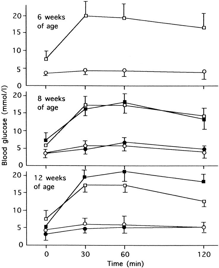
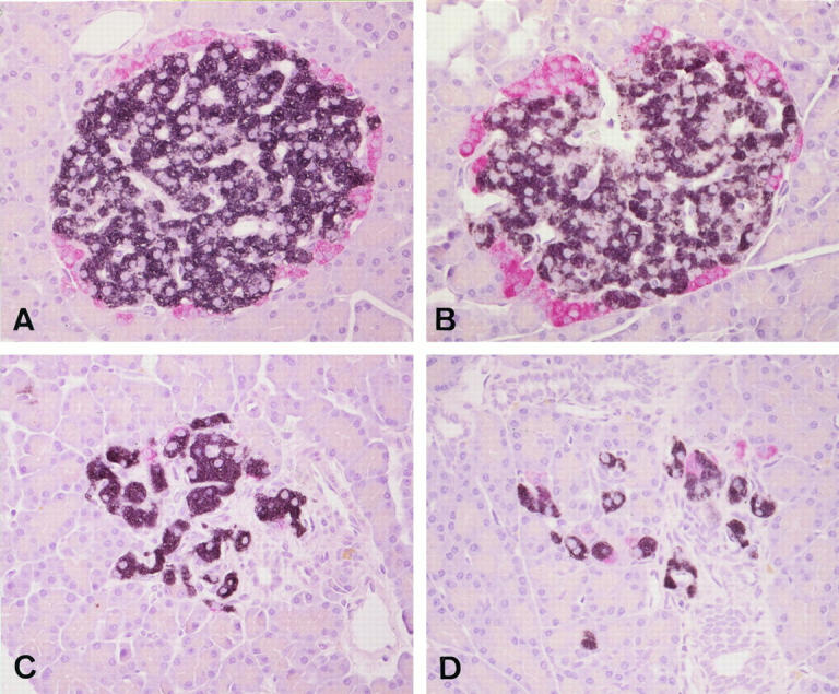
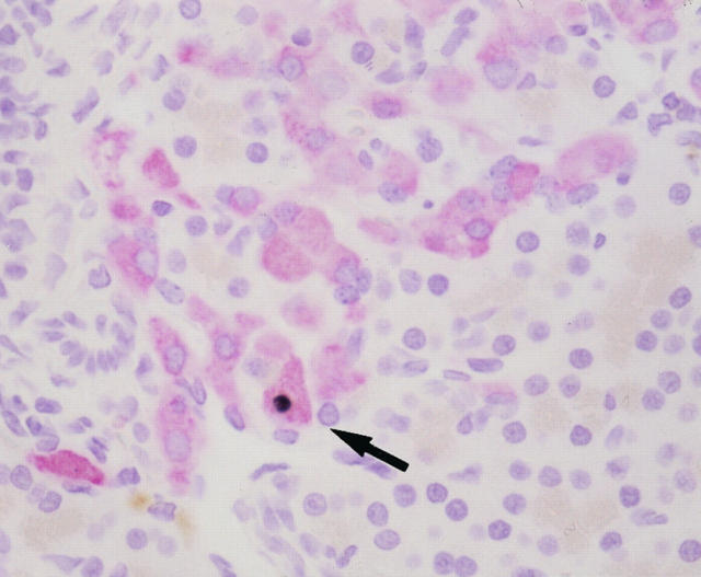
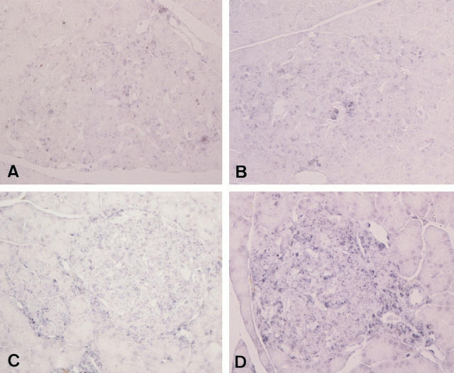
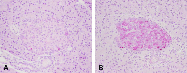
The Goto-Kakizaki (GK) rat is a spontaneously diabetic animal model of non-insulin-dependent diabetes mellitus, which is characterized by progressive loss of beta cells in the pancreatic islets with fibrosis. In the present study, we examined the effects of sucrose feeding on the islet pathology in this model. Six-week-old GK rats were fed with 30% sucrose for 6 weeks to induce severe hyperglycemia, and their condition was compared with that of nontreated rats. Age-matched normal Wistar rats were also given sucrose for the same periods and used for comparison. The sucrose-treated GK rats showed elevated blood glucose levels on oral glucose tolerance tests at 60 minutes and 120 minutes, representing 123% and 127% of values in untreated GK rats, respectively. At the end of the study, the mean beta-cell volume density in GK rats was 50% less than that in untreated Wistar rats. Sucrose feeding further reduced the volume densities of beta cells to only 50% of the levels of age-matched GK rats. Apoptotic cells were found in islet beta cells only in GK rats fed sucrose (mean 0.067%). There appeared to be more islets that immunohistochemically stained strongly positive with 8-hydroxy-deoxyguanosine as a marker of oxidative damage of DNA in GK rats fed sucrose compared with those not given sucrose. GK rats not fed sucrose showed significantly lower proliferative activity of beta cells measured by 5-bromo-2'-deoxyuridine uptake and intensified expression of Bcl-2 immunoreactivities at 6 weeks of age compared with those in age-matched Wistar rats. These two indices were reduced in both GK and Wistar rats with increasing age and were not affected by sucrose feeding in either group. The present study thus indicated that sucrose feeding promoted the apoptosis of beta cells in GK rats through increased oxidative stress without altering their proliferative activity.
Figures
References
-
- Weir GC, Leahy JL: Pathogenesis of non-insulin-dependent (type II) diabetes mellitus. ed 13 Kahn CR Weir GC eds. Joslin’s Diabetes Mellitus, 1994, :pp 240-263 Lea & Febiger, Philadelphia
-
- Cooppan R: General approach to the treatment of diabetes. ed 13 Kahn CR Weir GC eds. Joslin’s Diabetes Mellitus, 1994, :pp 397-403 Lea & Febiger, Philadelphia
-
- Clark A, Wells CA, Buley ID, Cruickshank JK, Vanhegan RI, Matthews DR, Cooper GJS, Holman RR, Turner RC: Islet amyloid, increased A-cells, reduced B-cells and exocrine fibrosis: quantitative changes in the pancreas in type 2 diabetes. Diabetes Res 1988, 9:151-159 - PubMed
-
- Saito K, Yaginuma N, Takahashi T: Differential volumetry of A, B and D cells in the pancreatic islets of diabetic and nondiabetic subjects. Tohoku J Exp Med 1979, 129:273-283 - PubMed
-
- Rahier J, Goebbels RM, Henquin JC: Cellular composition of the human diabetes pancreas. Diabetologia 1983, 24:366-371 - PubMed
MeSH terms
Substances
LinkOut - more resources
Full Text Sources
Medical
