. 1998 Aug;153(2):599-610.
doi: 10.1016/S0002-9440(10)65602-7.

Induction of nitric oxide synthase and microglial responses precede selective cell death induced by chronic impairment of oxidative metabolism

Affiliations

Induction of nitric oxide synthase and microglial responses precede selective cell death induced by chronic impairment of oxidative metabolism

N Y Calingasan et al. Am J Pathol. 1998 Aug.

Abstract

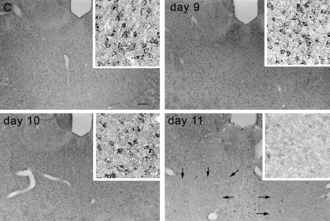
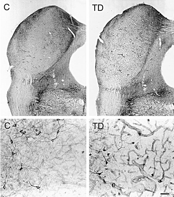
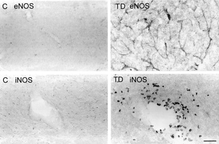
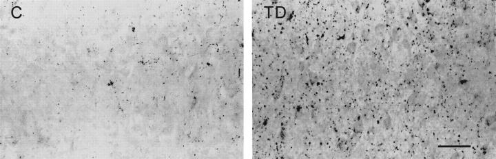
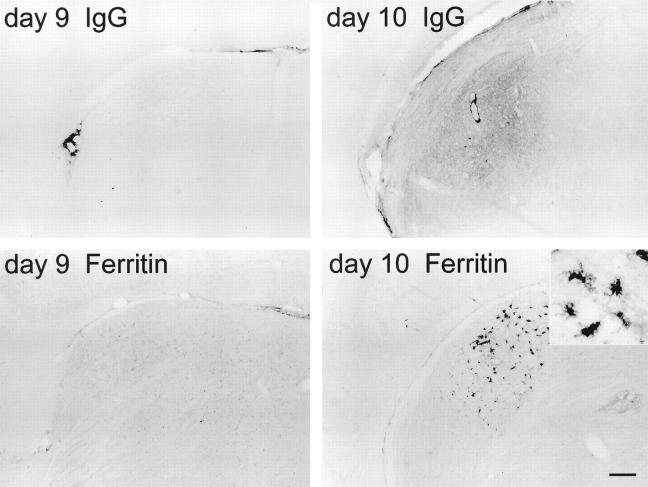
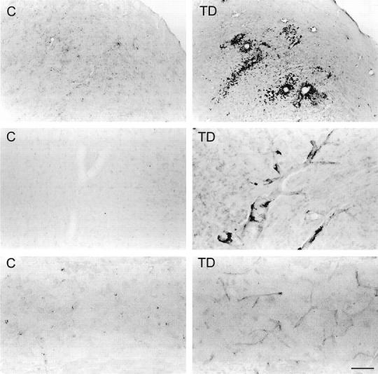
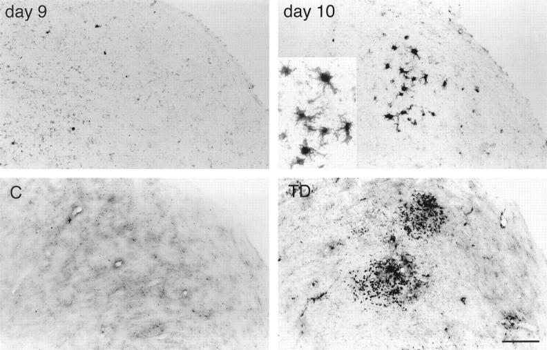
Abnormal oxidative processes including a reduction in thiamine-dependent enzymes accompany many neurodegenerative diseases. Thiamine deficiency (TD) models the cellular and molecular mechanisms by which chronic oxidative aberrations associated with thiamine-dependent enzyme deficits cause selective neurodegeneration. The mechanisms underlying selective cell death in TD are unknown. In rodent TD, the earliest region-specific pathological change is breakdown of the blood-brain barrier (BBB). The current studies tested whether nitric oxide and microglia are important in the initial events that couple BBB breakdown to selective neuronal loss. Enhanced expression of endothelial nitric oxide synthase and nicotinamide adenine dinucleotide phosphate diaphorase reactivity in microvessels, as well as the presence of numerous inducible nitric oxide synthase-immunoreactive microglia, accompanied the increases in BBB permeability. Nitric oxide synthase induction appears critical to TD pathology, because immunoreactivity for nitrotyrosine, a specific nitration product of peroxynitrite, also increased in axons of susceptible regions. In addition, TD elevated iron and the antioxidant protein ferritin in microvessels and in activated microglia, suggesting that these cells are responding to an oxidative challenge. All of these changes occurred in selectively vulnerable regions, preceding neuronal death. These findings are consistent with the hypothesis that the free radical-mediated BBB alterations permit entry of iron and extraneuronal proteins that set in motion a cascade of inflammatory responses culminating in selective neuronal loss. Thus, the TD model should help elucidate the relationship between oxidative deficits, BBB abnormalities, the inflammatory response, ferritin and iron elevation, and selective neurodegeneration.


Figures

Figure 1.
Figure 1.
βPP immunoreactivity in the mouse thalamus of control (C) and days 9, 10, and 11 of TD, demonstrating areas of neuronal loss at day 11 (arrows). Insets: High magnification of the mediodorsal thalamic nuclei. Scale bar = 100 μm (15 μm for insets).
Figure 2.
Figure 2.
Low-magnification (top) and high-magnification (bottom) photomicrographs showing NADPH-d staining of the inferior colliculus of a 12-day thiamine-deficient (TD) rat compared with control (C). Note the enhanced NADPH-d reactivity of the microvessels after TD. NADPH-d-stained neurons occur along the periphery (bottom, left side of each photomicrograph) of the vulnerable central nucleus of the inferior colliculus that contains virtually no stained neurons (bottom, right side of each photomicrograph). Scale bar = 100 μm (top) and 10 μm (bottom).
Figure 3.
Figure 3.
Endothelial NOS (eNOS) immunoreactivity (top) in microvessel walls within the thalamus of control (C) and thiamine-deficient (TD) mice depicting increased immunostaining after 10 days of TD. Inducible NOS (iNOS) immunodetection (bottom) in the thalamus of control (C) and thiamine-deficient (TD) mice shows immunoreactive macrophages around a blood vessel in the thalamus of TD. Scale bar = 100 μm (top) and 50 μm (bottom).
Figure 4.
Figure 4.
Immunocytochemical detection of nitrotyrosine formation in the paraventricular thalamic nucleus of control (C) and thiamine-deficient (TD) mice. Prominent immunostaining occurs in axons after thiamine deprivation. Scale bar = 25 μm.
Figure 5.
Figure 5.
IgG (top) and ferritin (bottom) immunoreactivities in the dorsal lateral thalamic nucleus of 9-day and 10-day thiamine-deficient mice. Accumulation of IgG immunoreactivity on day 10 parallels ferritin-labeled microglial activation. Inset: Morphology of typical activated microglia. Scale bar = 100 μm (25 μm for inset).
Figure 6.
Figure 6.
Top: Photomicrographs showing ferritin immunostaining in the inferior colliculus of control (C) and thiamine-deficient (TD) rats. Intensely labeled microglia occur prominently around blood vessels and are also scattered in the area of cell damage. Middle: Photomicrographs of ferritin in the thalamus of control and thiamine-deficient rats showing enhanced staining of large blood vessel walls in TD. Intensely labeled microglia occur along the walls of large vessels in TD. Bottom: Photomicrographs showing enhanced ferritin staining of capillaries in thiamine-deficient rat thalamus as compared with control. Scale bar = 250 μm (top) and 50 μm (middle and bottom).
Figure 7.
Figure 7.
Iron histochemical staining of mouse control (C) and thiamine-deficient (TD) lateral dorsal geniculate nucleus (top) and rat control and thiamine-deficient inferior colliculus (bottom). In TD, iron accumulates in microglia with a similar distribution as ferritin (see Figures 5 ▶ , bottom, and 6 ▶ , top). Scale bar = 50 μm (25 μm for inset).

Similar articles

Cited by

References

    1. Blass JP, Gibson GE: The role of oxidative abnormalities in the pathophysiology of Alzheimer’s disease. Rev Neurol (Paris) 1991, 147:513. - PubMed
    1. Smith CD, Carney JM, Starke-Reed PE, Oliver CN, Stadtman ER, Floyd RA, Markesbery WR: Excess brain protein oxidation and enzyme dysfunction in normal aging and in Alzheimer’s disease. Proc Natl Acad Sci USA 1991, 88:10540-10543 - PMC - PubMed
    1. Hensley K, Carney JM, Mattson MP, Aksenova M, Harris M, Wu JF, Floyd RA, Butterfield DA: A model for β-amyloid aggregation and neurotoxicity based on free radical generation by the peptide: relevance to Alzheimer’s Disease. Proc Natl Acad Sci USA 1994, 91:3270-3274 - PMC - PubMed
    1. Nixon RA, Cataldo AM: Free radicals, proteolysis and degeneration of neurons in Alzheimer’s disease: how essential is the β-amyloid link? Neurobiol Aging 1994, 15:463-469 - PubMed
    1. Smith MA, Perry G: Free radical damage, iron, and Alzheimer’s disease. J Neurol Sci 1995, 134:92-94 - PubMed

Publication types

MeSH terms

LinkOut - more resources