. 1998 Aug;153(2):639-48.
doi: 10.1016/S0002-9440(10)65605-2.

Hepatocellular hyperplasia, plasmacytoma formation, and extramedullary hematopoiesis in interleukin (IL)-6/soluble IL-6 receptor double-transgenic mice

Affiliations

Hepatocellular hyperplasia, plasmacytoma formation, and extramedullary hematopoiesis in interleukin (IL)-6/soluble IL-6 receptor double-transgenic mice

P Schirmacher et al. Am J Pathol. 1998 Aug.

Abstract

Cytokines interact not only with membrane anchored receptors, but also with specific soluble receptors which circulate in the bloodstream. In general, soluble cytokine receptors such as soluble tumor necrosis factor receptor, soluble interleukin 1 receptor, and soluble interleukin 4 receptor compete with their membrane-bound counterparts for the ligands and therefore act as antagonists. In contrast, soluble receptors for cytokines of the interleukin-6 (IL-6) family complex with their ligands act agonistically. Interestingly, the complex of IL-6 and the soluble interleukin 6 receptor (sIL-6R) activates target cells that do not express the membrane-bound IL-6R and therefore cannot respond to IL-6. To identify cellular responses that are due to IL-6/sIL-6R but not to IL-6 alone, IL-6/sIL-6R double-transgenic mice were generated and compared with IL-6 single-transgenic mice. IL-6/sIL-6R transgenic mice develop a severe phenotype showing 1) marked hepatocellular hyperplasia frequently surrounded by peliosis and necrosis, 2) significant acceleration and aggravation of plasmacytoma formation, and 3) excessive activation of extramedullary hematopoiesis in spleen and liver followed by a subsequent increase of all cellular components in the peripheral blood. These in vivo data suggest that the sIL-6R recruits primarily unresponsive cell populations such as hematopoietic progenitor cells and hepatocytes to IL-6-induced proliferation, but also enhances the known mitogenic effect of IL-6 on plasma cells and thereby contributes to plasmacytoma formation.


Figures
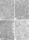
Figure 1.
Figure 1.
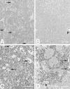
Accelerated development of plasmacytomas in IL-6/sIL-6R double-transgenic mice compared with IL-6 single transgenic mice. A: Focal periarteriolar plasma cell proliferation (arrows) in the spleen of an IL-6/sIL-6R double-transgenic mouse (7 weeks). Note the extensive activation of extramedullary hematopoiesis in the surrounding parenchyma. B: Normal spleen without plasma cell proliferation in an IL-6 transgenic mouse (8 weeks). Normal follicle (white pulp) is denoted by arrows. C: Plasmacytoma in the spleen of an IL-6/sIL-6R double-transgenic mouse (16 weeks). Note atypical plasma cells with eccentric round-to-ovoid pleomorphic and hyperchromatic nuclei (arrows). D: Excessive plasmacytoma in the spleen of an IL-6 single-transgenic mouse (16 weeks). Atypical plasma cells are marked by arrows. a in A and C denotes arterioles in the center of the white pulp. Scale bars, 100 μm.
Figure 2.
Figure 2.
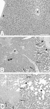
Extramedullary hematopoiesis in IL-6/sIL-6R double transgenic mice with splenic hematopoiesis preceding hepatic hematopoiesis. A: Highly activated diffuse extramedullary hematopoiesis in the spleen of IL-6/sIL-6R double-transgenic mice (6 weeks). Note the lack of definition between the red and white pulp (megakaryocytes indicated by arrows). B: Lack of extramedullary hematopoiesis in the liver of the same IL-6/sIL-6R double-transgenic mouse as shown in A (6 weeks) (p, portal tract). C: Excessive diffuse extramedullary hematopoiesis in the spleen of an IL-6/sIL-6R double-transgenic mouse (13 weeks). Some megakaryocytes are indicated by arrows. D: Extramedullary hematopoietic focus in a portal tract in the liver of the same IL-6/sIL-6R double-transgenic mouse as depicted in C (13 weeks). Bile ducts are labeled by arrows; bordering hepatic parenchyma is denoted by asterisk. Scale bars, 100 μm.
Figure 3.
Figure 3.
Time course of cellular blood composition in double- and single-transgenic mice as compared with control animals. A: White blood cell counts. B: Neutrophilic granulocyte counts. C: Thrombocyte counts. D: Red blood cell counts.
Figure 4.
Figure 4.
Severe liver pathology in IL-6/sIL-6R double-transgenic mice. A: Normal liver with regular trabecular arrangement of hepatocytes and lack of extramedullary hematopoiesis in an IL-6 single-transgenic animal (16 weeks). B: Overview of hepatic pathology in a 6-week-old IL-6/sIL-6R transgenic animal. Hepatocellular hyperplasia (marked by arrows) peripherally surrounded by peliosis and nonreactive necrosis (arrowheads). C: Extensive hepatic peliosis (*) in the vicinity of an areactive necrosis (arrows). Note the increased cellularity of liver cell plates as a sign of additional hepatocellular hyperplasia. (p, portal tracts; c, central vein). Scale bars, 100 μm.
Figure 5.
Figure 5.
Elevation of serum AST levels in IL-6/sIL-6R double-transgenic mice. Note the correlation of the necrosis score (+, mild; ++, moderate; +++, severe) with the increase of serum AST levels in double-transgenic mice.
Figure 6.
Figure 6.
Increased hepatocellular proliferation in IL-6/sIL-6R double-transgenic mice. C: Highly activated hepatocellular proliferation in an IL-6/sIL-6R double-transgenic mouse (7 weeks of age) with numerous PCNA-positive hepatocellular nuclei. By contrast, the number of PCNA-positive hepatocytes is low in a 7-week-old IL-6 single-transgenic mouse (A) and in a 7-week-old sIL-6R single-transgenic mouse (B). D: Graphic representation of the semiquantitative evaluation of hepatocellular proliferation in IL-6 single-transgenic mice (white bar), in sIL-6R single-transgenic mice (gray bar), and in IL-6/sIL-6R double-transgenic mice (black bar). Two animals were analyzed per strain. The percentage of PCNA-positive hepatocellular nuclei determined by evaluating 500 hepatocytes per animal ± standard deviation is presented in the figure.
Figure 7.
Figure 7.
Schematic chronology of pathology in IL-6/sIL-6R double-transgenic mice compared with IL-6 transgenic mice.

Similar articles

Cited by

References

    1. Akira S, Taga T, Kishimoto T: Interleukin-6 in biology and medicine. Adv Immunol 1993, 54:1-78 - PubMed
    1. Bauer J, Herrmann F: Interleukin-6 in clinical medicine. Ann Hematol 1991, 62:203-210 - PubMed
    1. Gauldie J, Richards C, Harnish D, Lansdorp P, Baumann H: Interferon beta 2/B-cell stimulatory factor type 2 shares identity with monocyte-derived hepatocyte-stimulating factor and regulates the major acute phase protein response in liver cells. Proc Natl Acad Sci USA 1987, 84:7251-7255 - PMC - PubMed
    1. Grötzinger J, Kurapkat G, Wollmer A, Kalai M, Rose-John S: The family of the IL-6-type cytokines: specificity and promiscuity of the receptor complexes. Proteins Struct Funct Genet 1997, 27:96-109 - PubMed
    1. Hibi M, Nakajima K, Hirano T: IL-6 cytokine family and signal transduction: a model of the cytokine system. J Mol Med 1996, 74:1-12 - PubMed

Publication types

MeSH terms