. 1998 Apr;57(4):231-6.
doi: 10.1136/ard.57.4.231.

Hydrostatic pressure induces expression of interleukin 6 and tumour necrosis factor alpha mRNAs in a chondrocyte-like cell line

Affiliations

Hydrostatic pressure induces expression of interleukin 6 and tumour necrosis factor alpha mRNAs in a chondrocyte-like cell line

K Takahashi et al. Ann Rheum Dis. 1998 Apr.

Abstract

Objectives: To clarify the effect of pressure on the expressions of proteoglycan core protein and metabolism related cytokines in a chondrocyte-like cell line, HCS-2/8.

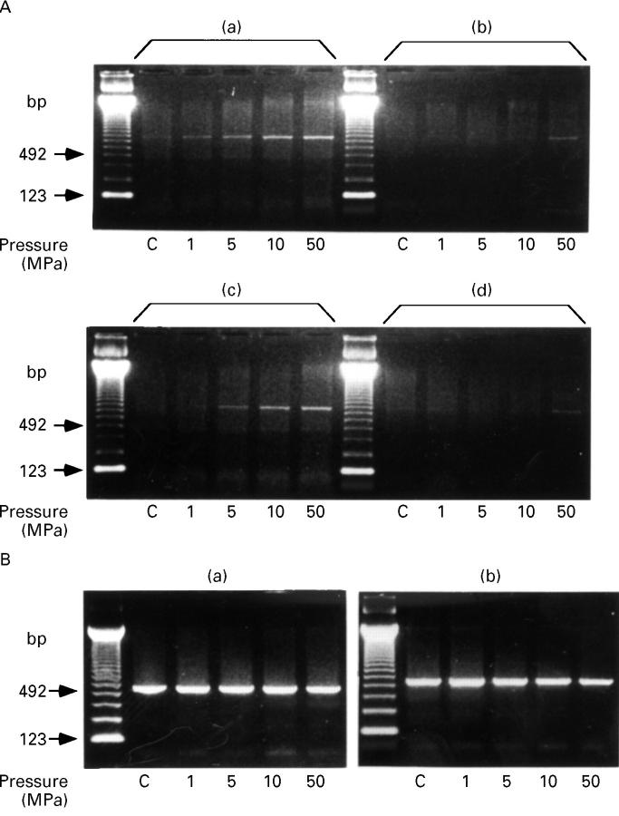
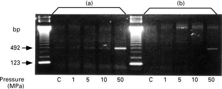
Methods: HCS-2/8 cells were exposed to 1, 5, 10, or 50 MPa of hydrostatic pressure (HP) for two hours, and mRNA expressions of interleukin 6 (IL6) and tumour necrosis factor alpha (TNFalpha) were examined by using reverse transcription-polymerase chain reaction (RT-PCR) method with specific primer sets; and mRNA of proteoglycan core protein, stromelysin, and tissue inhibitor of metalloproteinase 1 (TIMP1) were measured with northern blotting.

Results: HP exposure caused temporal morphological changes of the cells, but did not affect cellular viability, IL6 and TNFalpha mRNA expressions were not observed in the control cells under the atmospheric pressure, whereas in the cells treated with HP, pressure dependent enhancement of IL6 mRNA expression was observed between 30 minutes and four hours after HP release. TNFalpha mRNA expression also increased 30 minutes after the exposure to 50 MPa of HP and disappeared four hours later. Proteoglycan core protein mRNA levels increased between 30 minutes and four hours after the exposure to 1 or 5 MPa of HP, whereas the levels decreased after 10 or 50 MPa of HP. Stromelysin and TIMP1 mRNA signals did not respond to HP.

Conclusion: HP at excessively high levels induced IL6 and TNFalpha expression and reduced the expression of proteoglycan core protein, while physiological levels of HP increased the expression of proteoglycan core protein. These findings are important when considering the pathology of osteoarthritis.


Figures

Figure 1
Figure 1
Morphological changes of HCS-2/8 after exposure to HP. Phase contrast photomicrograms of cells cultured in DMEM containing 10% FBS after HP exposure. (A) Control cells under atmospheric pressure. (B) Thirty minutes after applying 50 MPa of HP. (C) Twenty four hours after applying 50 MPa of HP. (D) Thirty minutes after applying 5 MPa of HP. Bar = 100 µm.
Figure 2
Figure 2
Pressure dependent enhancement of IL6 mRNA expression. (A) IL6 RT-PCR products from the cells at 30 minutes (a) and (b) and four hours (c) and (d) after HP exposure. Two point five microgram (a) and (c) and 1 µg (b) and (d) of total RNA were reverse transcribed in 10 µl reaction mixture. (B) β actin RT-PCR products from the cells at 30 minutes (a) and two hours (b), which confirm cDNA isolation. One microgram of total RNA was reverse transcribed in 10 µl reaction mixture. C: control cells treated under the atmospheric pressure.
Figure 3
Figure 3
Induction of TNFα mRNA expression after HP exposure. The cells at 30 minutes after the exposure to 50 MPa of HP expressed TNFα mRNA. Two point five microgram (a) and 1 µg (b) of total RNA were reverse transcribed in 10 µl reaction mixture. No TNFα mRNA expression was detected in the cells between four and eight hours after HP exposure. C: control cells treated under the atmospheric pressure.
Figure 4
Figure 4
Effect of HP exposure on proteoglycan core protein mRNA expression. Cells were exposed to 1, 5, 10 or 50 MPa of HP for two hours, and total RNA was extracted at 30 minutes (A) and four hours (B) after the release of HP. Equal amounts of total RNA were fractionated by gel electrophoresis, transferred to a nylon membrane, and hybridised to RNA probes. For quantification, the signals were measured by using a densitometor. A photograph of gel staining (right side) shows equal amounts of RNA were applied. β actin mRNA expression is also shown. C: control cells treated under the atmospheric pressure.
Figure 5
Figure 5
Stromelysin and TIMP1 mRNA signals in HCS-2/8 cells after HP exposure. Representative signals at four hours after HP exposure are shown. C: control cells treated under the atmospheric pressure.

References

    1. Anal Biochem. 1987 Apr;162(1):156-9 - PubMed
    1. Proc Natl Acad Sci U S A. 1986 May;83(9):2879-83 - PubMed
    1. J Cell Sci Suppl. 1986;5:99-119 - PubMed
    1. EMBO J. 1987 Oct;6(10):2939-45 - PubMed
    1. Exp Cell Res. 1987 Jan;168(1):160-72 - PubMed

Publication types