

Articular cartilage superficial zone collagen birefringence reduced and cartilage thickness increased before surface fibrillation in experimental osteoarthritis
- PMID: 9709181
- PMCID: PMC1752579
- DOI: 10.1136/ard.57.4.237
Articular cartilage superficial zone collagen birefringence reduced and cartilage thickness increased before surface fibrillation in experimental osteoarthritis
Abstract
Objectives: To investigate articular cartilage collagen network, thickness of birefringent cartilage zones, and glycosaminoglycan concentration in macroscopically normal looking knee joint cartilage of young beagles subjected to experimental slowly progressive osteoarthritis (OA).
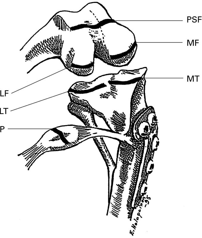
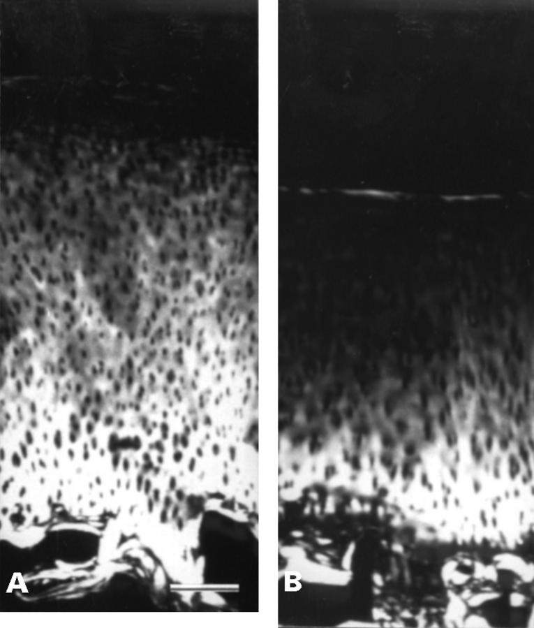
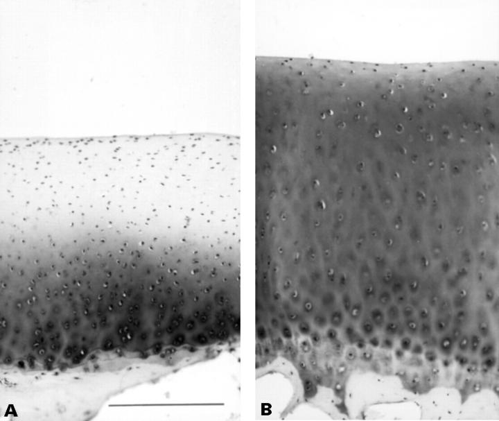
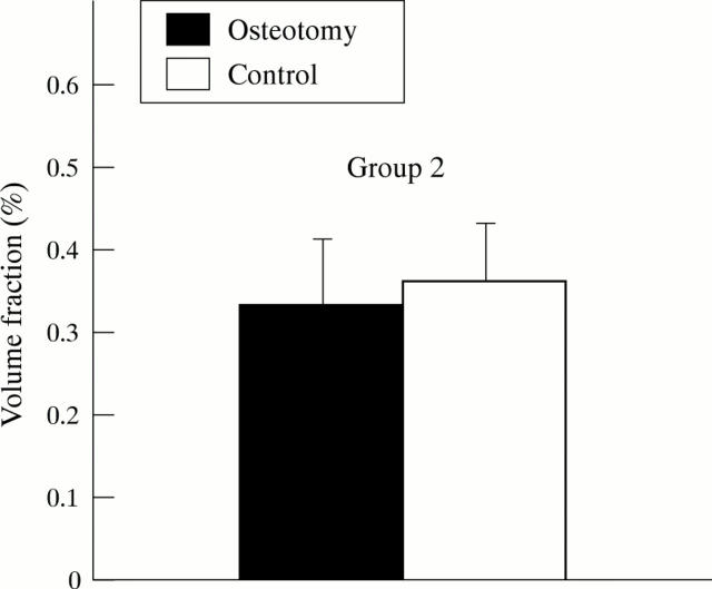
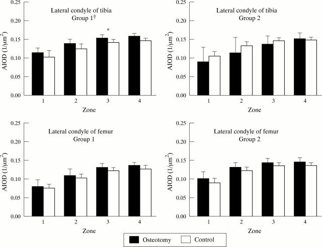
Methods: OA was induced by a tibial 30 degree valgus osteotomy in 15 female beagles at the age of 3 months. Fifteen sisters were controls. Cartilage specimens were collected seven (Group 1) and 18 months (Group 2) postoperatively. Collagen induced optical path difference and cartilage zone thickness measurements were determined from histological sections of articular cartilage with smooth and intact surface by computer assisted quantitative polarised light microscopy. Volume density of cartilage collagen fibrils was determined by image analysis from transmission electron micrographs and content of glycosaminoglycans by quantitative digital densitometry from histological sections.
Results: In the superficial zone of the lateral tibial and femoral cartilage, the collagen induced optical path difference (birefringence) decreased by 19 to 71% (p < 0.05) seven months postoperatively. This suggests that severe superficial collagen fibril network deterioration took place, as 18 months postoperatively, macroscopic and microscopic OA was present in many cartilage areas. Thickness of the uncalcified cartilage increased while the superficial zone became thinner in the same sites. In operated dogs, glycosaminoglycan content first increased (Group 1) in the lateral tibial condyle and then decreased (Group 2) (p < 0.05).
Conclusion: In this OA model, derangement of the superficial zone collagen network was the probable reason for birefringence reduction. This change occurred well before macroscopic OA.
Figures
Similar articles
-
Decreased birefringence of the superficial zone collagen network in the canine knee (stifle) articular cartilage after long distance running training, detected by quantitative polarised light microscopy.Ann Rheum Dis. 1996 Apr;55(4):253-64. doi: 10.1136/ard.55.4.253. Ann Rheum Dis. 1996. PMID: 8733443 Free PMC article.
-
Age matters: collagen birefringence of superficial articular cartilage is increased in young guinea-pigs but decreased in older animals after identical physiological type of joint loading.Osteoarthritis Cartilage. 2001 Nov;9(8):694-701. doi: 10.1053/joca.2001.0466. Osteoarthritis Cartilage. 2001. PMID: 11795988
-
Differences in submicroscopic structure of the extracellular matrix of canine femoral and tibial condylar articular cartilages as revealed by polarization microscopical analysis.Acta Biol Hung. 1996;47(1-4):341-53. Acta Biol Hung. 1996. PMID: 9124004
-
Relationship among biomechanical, biochemical, and cellular changes associated with osteoarthritis.Crit Rev Biomed Eng. 2001;29(4):373-91. doi: 10.1615/critrevbiomedeng.v29.i4.10. Crit Rev Biomed Eng. 2001. PMID: 11822479 Review.
-
[Comparative histomorphometry of subchondral bone density and articular cartilage thickness in the tibial head in early human arthritis].Z Orthop Ihre Grenzgeb. 1995 Jul-Aug;133(4):291-302. doi: 10.1055/s-2008-1039795. Z Orthop Ihre Grenzgeb. 1995. PMID: 7571794 Review. German.
Cited by
-
Polarized reflectance from articular cartilage depends upon superficial zone collagen network microstructure.Biomed Opt Express. 2019 Oct 3;10(11):5518-5534. doi: 10.1364/BOE.10.005518. eCollection 2019 Nov 1. Biomed Opt Express. 2019. PMID: 31799028 Free PMC article.
-
Macroscopic and radiological grading of osteoarthritis correlates inadequately with cartilage height and histologically demonstrable damage to cartilage structure.Rheumatol Int. 2005 Apr;25(3):161-8. doi: 10.1007/s00296-004-0582-6. Epub 2005 Feb 10. Rheumatol Int. 2005. PMID: 15703954
-
The effects of NSAIDs on types I, II, and III collagen metabolism in a rat osteoarthritis model.Rheumatol Int. 2012 Aug;32(8):2401-5. doi: 10.1007/s00296-011-1978-8. Epub 2011 Jun 17. Rheumatol Int. 2012. PMID: 21681568
-
In Situ Loading and Time-Resolved Synchrotron-Based Phase Contrast Tomography for the Mechanical Investigation of Connective Knee Tissues: A Proof-of-Concept Study.Adv Sci (Weinh). 2024 Jun;11(21):e2308811. doi: 10.1002/advs.202308811. Epub 2024 Mar 23. Adv Sci (Weinh). 2024. PMID: 38520713 Free PMC article.
-
Wnt7a Inhibits IL-1β Induced Catabolic Gene Expression and Prevents Articular Cartilage Damage in Experimental Osteoarthritis.Sci Rep. 2017 Feb 6;7:41823. doi: 10.1038/srep41823. Sci Rep. 2017. PMID: 28165497 Free PMC article.
References
Publication types
MeSH terms
Substances
LinkOut - more resources
Full Text Sources
Other Literature Sources
Medical
