. 1998 Sep;18(9):5099-108.
doi: 10.1128/MCB.18.9.5099.

Brachyury-related transcription factor Tbx2 and repression of the melanocyte-specific TRP-1 promoter

Affiliations

Brachyury-related transcription factor Tbx2 and repression of the melanocyte-specific TRP-1 promoter

S Carreira et al. Mol Cell Biol. 1998 Sep.

Abstract

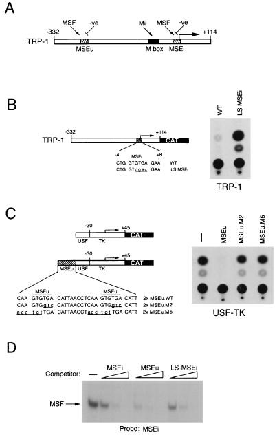
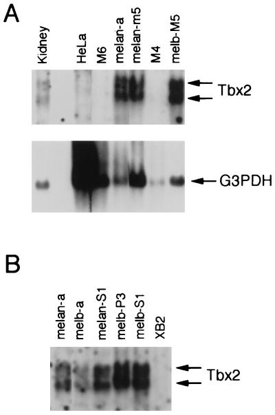
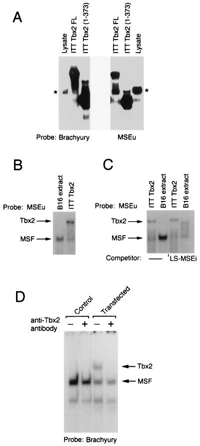
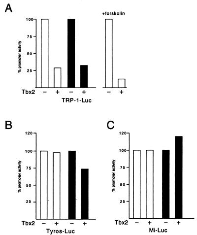
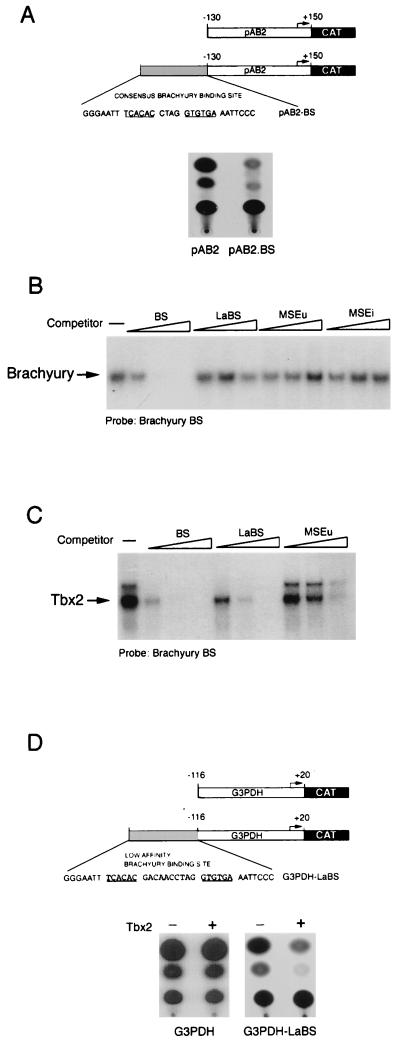
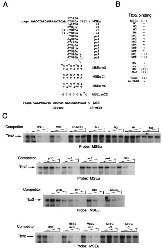
Previous work has demonstrated that two key melanocyte-specific elements termed the MSEu and MSEi play critical roles in the expression of the melanocyte-specific tyrosinase-related protein 1 (TRP-1) promoter. Both the MSEu and MSEi, located at position -237 and at the initiator, respectively, bind a melanocyte-specific factor termed MSF but are also recognized by a previously uncharacterized repressor, since mutations affecting either of these elements result in strong up-regulation of TRP-1 promoter activity in melanoma cells. Here we demonstrate that repression mediated by the MSEu and MSEi also operates in melanocytes. We also report that both the MSEu and MSEi are recognized by the brachyury-related transcription factor Tbx2, a member of the recently described T-box family, and that Tbx2 is expressed in melanocyte and melanoblast cell lines but not in melanoblast precursor cells. Although Tbx2 and MSF each recognize the TRP-1 MSEu and MSEi motifs, it is binding by Tbx-2, not binding by MSF, that correlates with repression. Several lines of evidence tend to point to the brachyury-related transcription factor Tbx2 as being the repressor of TRP-1 expression: both the MSEu and MSEi bind Tbx2, and mutations in either element that result in derepression of the TRP-1 promoter diminish binding by Tbx2; the TRP-1 promoter, but not the tyrosinase, microphthalmia, or glyceraldehyde-3-phosphate dehydrogenase (G3PDH) promoter, is repressed by Tbx2 in cotransfection assays; a high-affinity consensus brachyury/Tbx2-binding site is able to constitutively repress expression of the heterologous IE110 promoter; and a low-affinity brachyury/Tbx2 binding site is able to mediate Tbx2-dependent repression of the G3PDH promoter. Although we cannot rule out the presence of an additional, as yet unidentified factor playing a role in the negative regulation of TRP-1 in vivo, the evidence presented here suggests that Tbx2 most likely is the previously unidentified repressor of TRP-1 expression and as such is likely to represent the first example of transcriptional repression by a T-box family member.


Figures

FIG. 1
FIG. 1
Repression of TRP-1 in melanocytes is not mediated by MSF. (A) Schematic showing the elements required for regulation of the TRP-1 promoter. The M box is recognized by the transcription factor microphthalmia, while the MSEu and MSEi are targets for MSF and for negative regulation. (B) The LS-MSEi mutation relieves repression of TRP-1 in melanocytes. The melan-c melanocyte cell line was transfected with the indicated WT or LS-MSEi mutant TRP-1–CAT reporters, and CAT activity was determined. (C) Repression mediated by the MSEu in melanocytes. Melan-c cells were transfected with the USF-TK-CAT reporter or the indicated derivatives containing WT or mutant MSEu elements, and CAT activity was determined 48 h posttransfection. (D) MSF binds the WT and mutants LS-MSEi elements. A radioactive oligonucleotide probe containing the MSEu was used in a band shift assay with B16 melanoma cell nuclear extract, and MSF binding was competed with 10, 50, and 250 ng of unlabeled oligonucleotides containing the MSE, MSEu, or LS-MSEi. Only the bound DNA is shown. The sequence of each oligonucleotide probe and competitor is shown in Fig. 6.
FIG. 2
FIG. 2
The T-box transcription factor Tbx-2 is expressed in melanocytes and melanoblasts. (A) Northern blot of 5 μg of poly(A)+ mRNA derived from the indicated cell lines or from mouse kidney probed with either Tbx-2 or G3PDH cDNA. Tbx2 mRNA is present as two species of approximately 2.6 and 3.6 kb. The same blot was also probed for G3PDH message as a loading control. Note that the HeLa lane was deliberately overloaded to illustrate that these cells do not express Tbx2 message. M6 and M4 are premelanoblast cell lines; melan-a and melan-M5 are mouse melanocyte cell lines; melb-M5 is a mouse melanoblast cell line. (B) Northern blot of mRNA derived from the indicated cell lines and probed with radiolabeled Tbx-2 cDNA. Each lane was loaded with the same amount of RNA, as judged by staining with ethidium bromide and visualization under UV. melan-a and melan-S1 are mouse melanocyte cell lines; melb-a, melb-P3, and melb-S1 are mouse melanoblast cell lines; XB2 is a mouse keratinocyte cell line.
FIG. 3
FIG. 3
Tbx2 can bind the MSEu and MSEi and is distinct from MSF. (A) Band shift assay using a radiolabeled brachyury-binding site or the MSEu as the probe together with either unprogrammed reticulocyte lysate, ITT Tbx2FL, or the C-terminal truncation Tbx2(1-373). Only the bound DNA is shown. The asterisk indicates the position of a complex originating in the unprogrammed reticulocyte lysate and which has a mobility similar to that of MSF. (B) Band shift assay as in panel A, using the MSEu probe and either B16 nuclear extract or ITT Tbx2. The relative positions of the MSF- and Tbx2-containing complexes are indicated. (C) As for panel B but with 50 ng of the indicated LS-MSEi competitor where indicated. (D) Band shift assay using B16 extract derived from untransfected cells (control) or cells transfected with a Tbx2 expression vector, together with a brachyury consensus binding site as a probe. The DNA-binding reaction was performed in the presence or absence of a polyclonal anti-Tbx2 antiserum as indicated.
FIG. 4
FIG. 4
Specific repression of the TRP-1 promoter by Tbx2. B16 melanoma cells were transfected with the indicated TRP-1 (A), tyrosinase (Tyros) (B), or microphthalmia (Mi) (C) promoter-luciferase (Luc) reporters either alone or together with the Tbx2 expression vector. The open and filled columns represent the results from two independent transfection experiments. Forskolin was added at 20 μM to the indicated transfection assay. Luciferase activity was determined 48 h posttransfection.
FIG. 5
FIG. 5
Transcriptional repression by Tbx2 in melanocytes. (A) Schematic showing the structure of the herpes simplex virus IE110 promoter (in pAB2) and the consensus brachyury site (BS). (B) Brachyury does not bind the LaBS, the MSEu, or the MSEi, as determined by band shift assay using ITT brachyury together with the consensus brachyury probe and 10, 50, or 250 ng of the indicated consensus BS, LaBS, and MSEu, and MSEi competitors. (C) Tbx2 binds the consensus brachyury site around 5-fold better than the LaBS and around 15-fold better than the MSEu, as determined by band shift assay using ITT Tbx2 together with the consensus brachyury probe and 10, 50, or 250 ng of the consensus (BS) or low-affinity (LaBS) brachyury site or MSEu as the competitor. Only the bound DNA is shown. The full sequences of the BS and LaBS sites are shown in panels A and D, respectively. (D) Transcriptional repression by Tbx2. The indicated G3PDH-CAT or G3PDH.LaBS-CAT reporters were transfected into the melanocyte cell line melan-c either alone or together with a Tbx2 expression vector, and CAT activity was determined 48 h posttransfection.
FIG. 6
FIG. 6
DNA-binding specificity of Tbx2. A series of WT and mutant oligonucleotides based on the MSEu or MSEi was used in band shift assays (C) together with ITT Tbx2. The probes and competitors are indicated. Competitors were used at 10, 50, and 250 ng. The sequences or the probes and competitors are shown in panel A along with the full sequence of the parental MSEu or MSEi oligonucleotide. The derivatives used in the competition assays are identical except for the indicated residues shown in lowercase. mG, methylated G residue; mC, methylated C residue; I, inosine. For ease of reference, the bases within the GTGTGA motif are numbered 1 to 6. (B) Summary of the binding assays shown in panel C. Only the bound DNA is shown.

References

    1. Alexander M C, Lomanto M, Nasrin N, Ramaika C. Insulin stimulates glyceraldehyde-3-phosphate dehydrogenase gene expression through cis-acting DNA sequences. Proc Natl Acad Sci USA. 1988;85:5092–5096. - PMC - PubMed
    1. Baldwin C T, Lipsky N R, Hoth C F, Cohen T, Mamuya W, Milunsky A. Mutations in PAX3 associated with Waardenburg syndrome type I. Hum Mutat. 1994;3:205–211. - PubMed
    1. Bassi M T, Incerti B, Easty D J, Sviderskaya E V, Ballabio A. Cloning of the murine homolog of the ocular albinism type 1 (OA1) gene: sequence, genomic structure, and expression analysis in pigment cells. Genome Res. 1996;6:880–885. - PubMed
    1. Baynash A G, Hosoda K, Giaid A, Richardson J A, Emoto N, Hammer R E, Yanagisawa M. Interaction of endothelin-3 with endothelin-B receptor is essential for development of epidermal melanocytes and enteric neurons. Cell. 1994;79:1277–1285. - PubMed
    1. Bennett, D. Personal communication.

Publication types

MeSH terms