. 1998 Sep;18(9):5178-88.
doi: 10.1128/MCB.18.9.5178.

Tumor promoter arsenite activates extracellular signal-regulated kinase through a signaling pathway mediated by epidermal growth factor receptor and Shc

Affiliations

Tumor promoter arsenite activates extracellular signal-regulated kinase through a signaling pathway mediated by epidermal growth factor receptor and Shc

W Chen et al. Mol Cell Biol. 1998 Sep.

Abstract

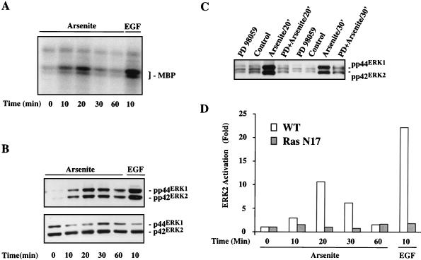
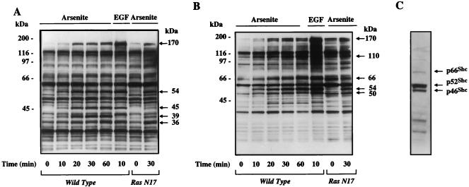
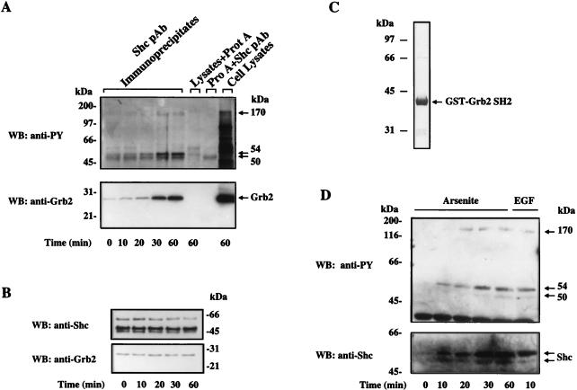
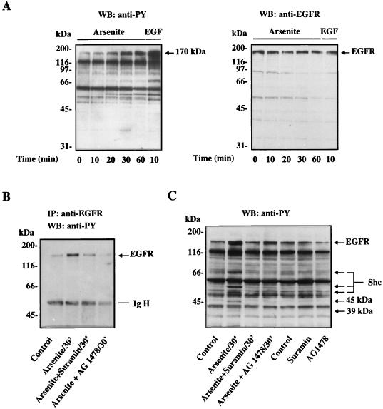
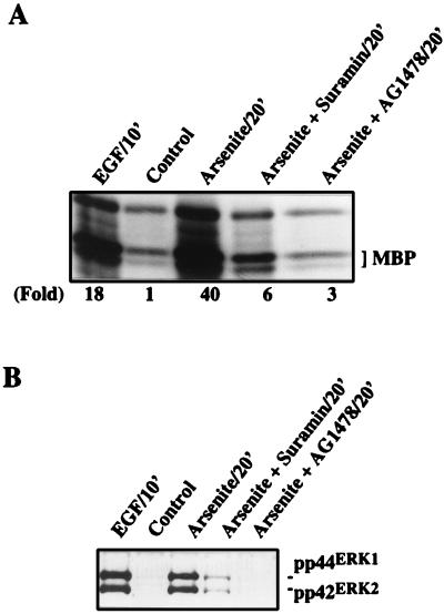
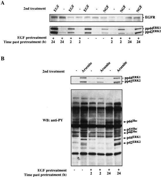
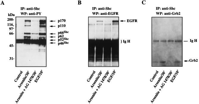
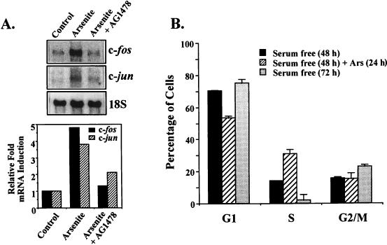
Although arsenite is an established carcinogen, the mechanisms underlying its tumor-promoting properties are poorly understood. Previously, we reported that arsenite treatment leads to the activation of the extracellular signal-regulated kinase (ERK) in rat PC12 cells through a Ras-dependent pathway. To identify potential mediators of the upstream signaling cascade, we examined the tyrosine phosphorylation profile in cells exposed to arsenite. Arsenite treatment rapidly stimulated tyrosine phosphorylation of several proteins in a Ras-independent manner, with a pattern similar to that seen in response to epidermal growth factor (EGF) treatment. Among these phosphorylated proteins were three isoforms of the proto-oncoprotein Shc as well as the EGF receptor (EGFR). Tyrosine phosphorylation of Shc allowed for enhanced interactions between Shc and Grb2 as identified by coimmunoprecipitation experiments. The arsenite-induced tyrosine phosphorylation of Shc, enhancement of Shc and Grb2 interactions, and activation of ERK were all drastically reduced by treatment of cells with either the general growth factor receptor poison suramin or the EGFR-selective inhibitor tyrphostin AG1478. Down-regulation of EGFR expression through pretreatment of cells with EGF also attenuated ERK activation and Shc tyrosine phosphorylation in response to arsenite treatment. These results demonstrate that the EGFR and Shc are critical mediators in the activation of the Ras/ERK signaling cascade by arsenite and suggest that arsenite acts as a tumor promoter largely by usurping this growth factor signaling pathway.


Figures

FIG. 1
FIG. 1
Arsenite-stimulated ERK activation depends on upstream signals. Serum-starved PC12 cells were treated with either 400 μM arsenite or 100 ng of EGF per ml for the indicated times (in all figures, ′ denotes minutes). (A) Time course of ERK activation following arsenite treatment of PC12 cells. Kinase activity of ERK was measured by an immune complex kinase assay using MBP as a substrate. (B) Western blot analysis of phosphorylated and total ERK protein in arsenite-treated cells. Upper panel, phospho-ERK detected with an anti-phospho-ERK antibody; lower panel, total ERK levels detected with an anti-pan-ERK antibody. (C) Effect of MEK inhibitor PD98059 on phosphorylation of ERK in response to arsenite. Cells were preincubated with 50 μM PD98059 for 30 min before addition of arsenite to the medium. Phospho-ERK was detected by Western blot analysis using the phospho-ERK-specific antibody. (D) Comparison of ERK kinase activities in wild-type (WT) and Ras N17 mutant PC12 cells. ERK activity was assessed by an immune complex kinase assay using MBP as a substrate and quantitated by using ImageQuant software from Molecular Dynamics (Sunnyvale, Calif.). Results are representative of three separate experiments.
FIG. 2
FIG. 2
Protein tyrosine phosphorylation profile of arsenite-treated PC12 cells. (A) Wild-type and Ras N17 PC12 cells were treated with 400 μM arsenite or 100 ng of EGF per ml for the indicated times. The cell extracts were separated on 10% NuPAGE Bis-Tris gel. Tyrosine-phosphorylated proteins were detected by immunoblotting with the antiphosphotyrosine antibody 4G10. The major tyrosine-phosphorylated bands induced by arsenite or EGF are indicated by arrows. (B) Protein tyrosine phosphorylation profile in arsenite-treated PC12 cells was generated on a 4 to 12% gradient NuPAGE Bis-Tris gel. Tyrosine-phosphorylated proteins were detected by immunoblotting with the antiphosphotyrosine antibody 4G10. Arrows indicate the major tyrosine-phosphorylated bands induced by arsenite or EGF. (C) Shc isoforms comigrate with tyrosine-phosphorylated proteins induced by arsenite or EGF. The blot shown in panel B was stripped and reprobed with an Shc-specific rabbit polyclonal antibody. Shown is a single lane from this blot (wild-type, arsenite-treated cells), as all lanes showed identical patterns.
FIG. 3
FIG. 3
Arsenite stimulates Shc tyrosine phosphorylation and its interaction with Grb2. (A) Upper panel, arsenite-induced tyrosine phosphorylation of Shc. Cellular extracts from arsenite-treated PC12 cells were immunoprecipitated with anti-Shc polyclonal antibody (pAb) and protein (Prot or Pro) A-Sepharose beads. Shc immunoprecipitates were resolved by NuPAGE Bis-Tris gel and Western blotted (WB) with the antiphosphotyrosine (anti-PY) antibody 4G10. Cell lysates (60-min arsenite treatment) and immunoprecipitates obtained in the absence of either Shc antibody or lysates were run as controls. Arrows indicate the major tyrosine-phosphorylated bands detected. Lower panel, arsenite-enhanced interaction between Grb2 and Shc. The immunoblot used in the upper panel was stripped and then blotted with a monoclonal anti-Grb2 antibody. The position of Grb2 is indicated. (B) Western blot analysis of total Shc and Grb2 levels in control and arsenite-treated cells. (C) Coomassie blue staining of the GST-Grb2 SH2 domain fusion protein. Recombinant GST-Grb2 SH2 domain fusion protein was produced in E. coli and purified by glutathione-Sepharose affinity column followed by FPLC. The fusion protein was then separated by SDS-polyacrylamide gel electrophoresis and stained. (D) Arsenite stimulates interaction between Grb2 SH2 domain and Shc in vitro. Cell lysates prepared from arsenite- or EGF-treated cells were incubated with the glutathione-free GST-Grb2 SH2 fusion protein for 2 h at 4°C. GST-Grb2 SH2 domain fusion protein and proteins associated with it were recovered by glutathione-Sepharose affinity chromatography and subjected to immunoblotting with the antiphosphotyrosine 4G10 or the anti-Shc antibodies.
FIG. 4
FIG. 4
Role of EGFR in mediating arsenite-induced protein tyrosine phosphorylation. (A) Control and arsenite-treated PC12 cells were subjected to Western blot (WB) analysis using the antiphosphotyrosine (anti-PY) monoclonal antibody 4G10 and an anti-EGFR polyclonal antibody. The same blot was sequentially probed with the two antibodies. The band recognized by the EGFR antibody is indicated by an arrow. EGFR detected in the right panel comigrates with the 170-kDa tyrosine-phosphorylated protein seen in the left panel. (B) Effects of suramin and tyrphostin AG1478 on the tyrosine phosphorylation of EGFR. Suramin (300 μM) or tyrphostin AG1478 (40 nM) was added at the same time as arsenite (400 μM). Cell lysates were immunoprecipitated (IP) with the anti-EGFR polyclonal antibody, and the immunoprecipitates were subjected to Western blot analysis using the antiphosphotyrosine antibody 4G10. (C) Effect of suramin and tyrphostin AG1478 on overall arsenite-induced protein tyrosine phosphorylation. PC12 cells were treated with arsenite in the presence or absence of suramin or tyrphostin AG1478 under the conditions described above. Cell lysates were analyzed by Western blotting with the antiphosphotyrosine antibody 4G10. Arrows denote proteins whose tyrosine phosphorylation was affected by suramin or tyrphostin AG1478.
FIG. 5
FIG. 5
Central role of EGFR in mediating arsenite-induced ERK activation. (A) Effects of suramin and tyrphostin AG1478 on arsenite-induced ERK activation. PC12 cells were treated with arsenite (400 μM) in the presence or absence of 300 μM suramin or 40 nM tyrphostin AG1478 for 30 min. ERK activity was analyzed by an immune complex kinase assay using MBP as a substrate and quantitated by using ImageQuant software (Molecular Dynamics). (B) Effects of suramin and tyrphostin AG1478 on arsenite-induced ERK phosphorylation. Cell lysates from cells treated with arsenite in the absence or presence of suramin or tyrphostin AG1478 were analyzed by Western blotting using the phospho-ERK-specific antibody.
FIG. 6
FIG. 6
Pretreatment of cells with EGF inhibits a subsequent arsenite response. (A) PC12 cells were pretreated with EGF (1 μg/ml) for 1 h (+) or not pretreated (−), as indicated at the bottom. The cells were then washed and incubated in EGF-free medium for the time intervals shown at the bottom. The cells were further treated with EGF (100 ng/ml) for 10 min or NGF (100 ng/ml) for 5 min (second treatment, shown at the top). Cell lysates following these different treatments were subjected to Western blot analysis using anti-EGFR polyclonal antibody (upper panel). The same blot was sequentially probed with anti-phospho-ERK-specific antibody (lower panel). (B) PC12 cells were pretreated with EGF (1 μg/ml) for 1 h (+) or not pretreated (−), as indicated at the bottom. The cells were then washed and incubated in EGF-free medium for the time intervals shown at the bottom. The cells were further treated (second treatment) with 400 μM arsenite for an additional 30 min. Total cellular proteins were subjected to Western blot (WB) analysis using anti-phospho-ERK-specific antibody (upper panel) or the antiphosphotyrosine (anti-PY) monoclonal antibody 4G10 (lower panel). EGFR-mediated protein tyrosine phosphorylation stimulated by arsenite is indicated by arrows.
FIG. 7
FIG. 7
Treatment of PC12 cells with the EGFR-selective inhibitor tyrphostin AG1478 prevents arsenite-induced Shc tyrosine phosphorylation and its interaction with EGFR and Grb2. PC12 cells were treated with EGF or arsenite in the presence or absence of 30 nM tyrphostin AG1478. Cell lysates containing 4.5 mg of protein were subjected to immunoprecipitation (IP) by using 1 μg of anti-Shc polyclonal antibody. Immunoprecipitates were subjected to Western blot (WB) analysis using the antiphosphotyrosine (anti-PY) antibody 4G10 (A), anti-EGFR antibody (B), and anti-Grb2 antibody (C). The same blot was sequentially probed with the three antibodies.
FIG. 8
FIG. 8
Arsenite induces c-fos and c-jun expression and enhances DNA synthesis. (A) Northern blot analysis of RNA extracted from arsenite-stimulated PC12 cells in the presence or absence of tyrphostin AG1478. PC12 cells were treated (Arsenite) or untreated (Control) with 400 μM arsenite for 30 min in the presence (Arsenite + AG1478) or absence of tyrphostin AG1478. The cells were harvested and analyzed for expression of c-fos mRNA, c-jun mRNA, and 18S rRNA by Northern blot analysis. The signals were quantitated with a PhosphorImager, and following normalization to 18S rRNA, relative fold mRNA induction for c-fos and c-jun mRNA expression was determined (bottom panel). (B) Arsenite increases cell population in S phase. PC12 cells were starved in serum-free medium for 48 h. Starved cells were either treated with 400 μM arsenite for 24 h or continual kept in serum-free medium for another 24 h. For cell cycle distribution analysis (as described in Materials and Methods), the cells were collected before arsenite treatment [Serum free (48 h)], after arsenite treatment [Serum free (48 h) + Ars (24 h)], or without treatment [serum free (72 h)]. The percentages of the cells in various cell cycle phases are indicated as means ± standard deviations. Results were from three different experiments.

References

    1. Angel P, Karin M. The role of Jun, Fos and the AP-1 complex in cell-proliferation and transformation. Biochim Biophys Acta. 1991;1072:129–157. - PubMed
    1. Avruch J, Zhang X F, Kyriakis J M. Raf meets Ras: completing the framework of a signal transduction pathway. Trends Biochem Sci. 1994;19:279–283. - PubMed
    1. Bagla P, Kaiser J. India’s spreading health crisis draws global arsenic experts. Science. 1996;274:174–175. - PubMed
    1. Bonfini L, Migliaccio E, Pelicci G, Lanfrancone L, Pelicci P G. Not all Shc’s roads lead to Ras. Trends Biochem Sci. 1996;21:257–261. - PubMed
    1. Carpenter G, Cohen S. 125I-labeled human epidermal growth factor. Binding, internalization, and degradation in human fibroblasts. J Cell Biol. 1976;71:159–171. - PMC - PubMed

MeSH terms