. 1998 Sep;18(9):5189-98.
doi: 10.1128/MCB.18.9.5189.

Genetic, physical, and functional interactions between the triphosphatase and guanylyltransferase components of the yeast mRNA capping apparatus

Affiliations

Genetic, physical, and functional interactions between the triphosphatase and guanylyltransferase components of the yeast mRNA capping apparatus

C K Ho et al. Mol Cell Biol. 1998 Sep.

Abstract

We have characterized an essential Saccharomyces cerevisiae gene, CES5, that when present in high copy, suppresses the temperature-sensitive growth defect caused by the ceg1-25 mutation of the yeast mRNA guanylyltransferase (capping enzyme). CES5 is identical to CET1, which encodes the RNA triphosphatase component of the yeast capping apparatus. Purified recombinant Cet1 catalyzes hydrolysis of the gamma phosphate of triphosphate-terminated RNA at a rate of 1 s-1. Cet1 is a monomer in solution; it binds with recombinant Ceg1 in vitro to form a Cet1-Ceg1 heterodimer. The interaction of Cet1 with Ceg1 elicits >10-fold stimulation of the guanylyltransferase activity of Ceg1. This stimulation is the result of increased affinity for the GTP substrate. A truncated protein, Cet1(201-549), has RNA triphosphatase activity, heterodimerizes with and stimulates Ceg1 in vitro, and suffices when expressed in single copy for cell growth in vivo. The more extensively truncated derivative Cet1(246-549) also has RNA triphosphatase activity but fails to stimulate Ceg1 in vitro and is lethal when expressed in single copy in vivo. These data suggest that the Cet1-Ceg1 interaction is essential but do not resolve whether the triphosphatase activity is also necessary. The mammalian capping enzyme Mce1 (a bifunctional triphosphatase-guanylyltransferase) substitutes for Cet1 in vivo. A mutation of the triphosphatase active-site cysteine of Mce1 is lethal. Hence, an RNA triphosphatase activity is essential for eukaryotic cell growth. This work highlights the potential for regulating mRNA cap formation through protein-protein interactions.


Figures

FIG. 1
FIG. 1
CES5 is a multicopy suppressor of ceg1-25. ceg1-25 cells were transformed with a 2μ-URA3 plasmid containing the wild-type CEG1 gene, with the 2μ URA3 CES5 plasmid [pCES5(10.4)] that was isolated in the multicopy suppressor screen, with pCES5(2.6 kb) containing the YPL228W reading frame, and with the YEp24 vector plasmid without any insert. Ura+ transformants were selected and grown at 25°C in liquid culture in medium lacking uracil. The cultures were adjusted to an A600 of 0.1, and aliquots of serial 10-fold dilutions were spotted on agar medium lacking uracil. The plates were photographed after incubation for 3 days at either 25 or 37°C. aa, amino acids.
FIG. 2
FIG. 2
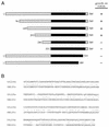
Deletion analysis. (A) CEN TRP1 plasmids encoding full-length and truncated versions of the YPL228W protein were transformed into YBS20. Individual Trp+ transformants were selected and patched to plates lacking tryptophan. Cells were streaked on plates containing 0.75-mg/ml 5-FOA. The plates were incubated at 25 and 30°C. Alleles that supported the formation of wild-type size colonies after 3 days on 5-FOA were scored as positive. Lethal truncation alleles (scored negative) were those that formed no colonies after 7 days at either temperature. The full-length and truncated polypeptides are depicted as horizontal bars with N termini at the left and C termini at the right. The region of homology between the YPL228W and YMR180C gene products is in black. (B) The YPL228W amino acid sequence between residues 302 and 532 is aligned with the homologous segment of the yeast YMR180C polypeptide (residues 84 to 310). Identical amino acids are indicated by colons, and conserved residues are denoted by periods. Discontinuities in the alignment are indicated by dashes.
FIG. 3
FIG. 3
Purification and RNA triphosphatase activity of Cet1. (A) The elution profile of the Cet1 polypeptide during phosphocellulose column chromatography was analyzed by SDS-PAGE. Lanes: Ni, Ni-agarose eluate fraction applied to the phosphocellulose column; F, phosphocellulose flowthrough fraction; W, 50 mM NaCl wash fraction; 0.1, 100 mM NaCl eluate; 0.2, 200 mM NaCl eluate; 0.5, 500 mM NaCl eluate; 1.0, 1.0 M NaCl eluate. A Coomassie blue-stained gel is shown. The values to the right are molecular sizes in kilodaltons. (B) RNA triphosphatase activity. Reaction mixtures (10 μl) containing 50 mM Tris HCl (pH 7.5), 5 mM DTT, 20 pmol (of triphosphate termini) of γ-32P-labeled poly(A), either 1 mM MgCl2 (+Mg) or no divalent cation (−Mg), and the indicated amounts of Cet1 (0.1 M NaCl phosphocellulose fraction) were incubated for 15 min at 30°C. Pi release is plotted as a function of input protein. (C) Magnesium titration. Reaction mixtures containing 20 pmol of γ-32P-labeled poly(A), 0.5 ng of Cet1, and MgCl2 as specified were incubated for 15 min at 30°C.
FIG. 4
FIG. 4
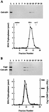
Analysis of Cet1-Ceg1 interaction by glycerol gradient sedimentation. Aliquots (0.2 ml) of protein samples were applied to 4.8-ml 15 to 30% glycerol gradients containing 50 mM Tris-HCl (pH 8.0), 0.1 M NaCl, 2 mM DTT, and 0.05% Triton X-100. The gradients were centrifuged at 50,000 rpm for 13 h at 4°C in a Beckman SW50 rotor. Fractions (∼0.21 ml) were collected from the bottom of the tube (fraction 1). Aliquots (25 μl) of alternate fractions were analyzed by SDS-PAGE along with an aliquot of the material that had been applied to the gradient (lane L). The gels were fixed and stained with Coomassie blue dye. The identities of the polypeptides are indicated. (A) Sedimentation of Cet1. A 10-μg sample of the phosphocellulose enzyme fraction was applied to the glycerol gradient. Gradient fractions were assayed for RNA triphosphatase (•). The RNA triphosphatase reaction mixtures contained 20 pmol of γ-32P-labeled poly(A) and 1 μl of a 1/50 dilution of the indicated gradient fractions. Incubation was for 15 min at 30°C. (B) Sedimentation of Ceg1. A 20-μg sample of the heparin agarose Ceg1 fraction was applied to the gradient. Gradient fractions were assayed for enzyme-GMP complex formation (○). The guanylyltransferase reaction mixtures contained 0.17 μM [α-32P]GTP and 1 μl of the indicated fractions. The reaction products were analyzed by SDS-PAGE, and the signal intensity of the radiolabeled Ceg1 polypeptide (PSL, photostimulatable luminescence) was measured by scanning the dried gel with a PhosphorImager. (C) Sedimentation of a mixture of Cet1 (10 μg) and Ceg1 (20 μg). The two proteins were mixed in buffer C containing 0.1 M NaCl and then incubated on ice for 30 min before being applied to the glycerol gradient. Gradient fractions were assayed for RNA triphosphatase (•) and enzyme-GMP complex formation (○) as specified for panels A and B. The peaks of the marker proteins catalase, bovine serum albumin (BSA), and cytochrome c (cyt C), which were centrifuged in a parallel gradient, are indicated.
FIG. 5
FIG. 5
Stimulation of Ceg1-GMP complex formation by Cet1 and Cet1(201-549). (A) Purification of Cet1(201-549) and Cet1(246-549). Aliquots (3 μg) of the phosphocellulose preparations of full-length Cet1 (lane 1), Cet1(201-549) (lane 2), and Cet1(246-549) (lane 3) were analyzed by SDS-PAGE. A Coomassie blue-stained gel is shown. The positions and sizes (in kilodaltons) of marker proteins are indicated on the left. (B) Effect of Cet1 on Ceg1 guanylyltransferase activity. Ceg1 (300 ng) was mixed with 300 ng of Cet1 (lane 2), Cet1(201-549) (lane 3), or Cet1(246-549) (lane 4) or with 320 ng of the mouse RNA triphosphatase domain Mce1(1-210) (lane 5) in 10 μl of buffer C containing 75 mM NaCl. A control sample contained 300 ng of Ceg1 alone (lane 1). The mixtures were incubated for 30 min on ice. An aliquot (1 μl) of each sample was then assayed for enzyme-GMP complex formation. Reaction mixtures (20 μl) containing 50 mM Tris HCl (pH 8.0), 5 mM MgCl2, 5 mM DTT, 0.17 μM [α-32P]GTP, and 1 μl of enzyme were incubated for 10 min at 37°C. The reaction products were analyzed by SDS-PAGE. Ceg1-GMP complex formation was visualized by autoradiography of the dried gel. (C) Cet1 titration. Ceg1 (550 ng) was mixed with 0, 65, 130, 325, 650, or 1,300 ng of Cet1 in 20 μl of buffer C containing 75 mM NaCl. An aliquot (1 μl) of each sample was then assayed for enzyme-GMP complex formation. Reaction mixtures (20 μl) contained 50 mM Tris HCl (pH 8.0), 5 mM MgCl2, 5 mM DTT, 0.17 μM [α-32P]GTP, and 28 ng of Ceg1 plus Cet1 as specified. Ceg1-[32P]GMP complex formation was quantitated by scanning the SDS-PAGE gel. The effect of Cet1 on Ceg1 activity (fold stimulation) was calculated as the ratio of the Ceg1-[32P]GMP signal intensity in Cet1-containing reaction mixtures to Ceg1-[32P]GMP signal intensity in the control reaction mixture lacking Cet1. The data shown are averages of two separate titration experiments. (D) GTP titration. Ceg1 (300 ng) was mixed with 300 ng of Cet1 in 10 μl of buffer C containing 75 mM NaCl (+ Cet1). A control sample contained 300 ng of Ceg1 alone (− Cet1). The mixtures were incubated for 30 min on ice. An aliquot (1 μl) of each sample was then assayed for enzyme-GMP complex formation in reaction mixtures (20 μl) containing 50 mM Tris HCl (pH 8.0), 5 mM MgCl2, 5 mM DTT, and [α-32P]GTP as specified. Incubation was for 10 min at 37°C. Ceg1-GMP complex formation is plotted as a function of GTP concentration.
FIG. 6
FIG. 6
Cet1(201-549) forms a complex with Ceg1. Aliquots (0.2 ml) of protein samples were applied to 4.8-ml 15 to 30% glycerol gradients containing 50 mM Tris-HCl (pH 8.0), 0.1 M NaCl, 2 mM DTT, and 0.05% Triton X-100. The gradients were centrifuged at 50,000 rpm for 16 h at 4°C in a Beckman SW50 rotor. Fractions (∼0.21 ml) were collected from the bottom of the tube (fraction 1). Aliquots (20 μl) of alternate fractions were analyzed by SDS-PAGE along with an aliquot of the material that had been applied to the gradient (lane L). The gels were fixed and stained with Coomassie blue dye. (A) Sedimentation analysis of Cet1(201-549). A 20-μg sample of the phosphocellulose preparation of Cet1(201-549) was applied to the gradient. Glycerol gradient fractions were assayed for RNA triphosphatase activity in reaction mixtures containing 20 pmol of γ-32P-labeled poly(A) and 1 μl of a 1/50 dilution of the indicated fractions. Incubation was for 15 min at 30°C. The peaks of the marker proteins catalase, bovine serum albumin (BSA), and cytochrome c (cyt C), which were centrifuged in a parallel gradient, are indicated. (B) Cet1(201-549) (20 μg) and Ceg1 (20 μg) were mixed in buffer C containing 0.1 M NaCl and then incubated on ice for 30 min before being applied to the glycerol gradient. Gradient fractions were assayed for RNA triphosphatase as specified for panel A. Guanylyltransferase reaction mixtures contained 0.17 μM [α-32P]GTP and 1 μl of the indicated fractions. PSL, photostimulable luminescence.
FIG. 7
FIG. 7
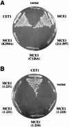
Complementation of Δcet1 by MCE1. (A) Δcet1 strain YBS20 was transformed with a CEN TRP1 plasmid bearing wild-type CET1, with pYX132(CEN TRP1)-based plasmids containing either wild-type MCE1 or mutant allele MCE1(K294A), MCE1(C126A), or MCE1(211-597), or with the pYX132 vector (7). (B) YBS20 was transformed with pYX132-based plasmid MCE1(1-231) or MCE1(1-210) (two independent isolates of each plasmid were tested), with the pYX132 vector, or with the CET1 control. Individual Trp+ transformants were selected and then patched on agar medium lacking tryptophan. Cells from single patches were then streaked on agar medium containing 0.75-mg/ml 5-FOA. The plates were photographed after incubation for 4 days at 30°C.

References

    1. Bi E, Pringle J R. ZDS1 and ZDS2, genes whose products may regulate Cdc42p in Saccharomyces cerevisiae. Mol Cell Biol. 1996;16:5264–5275. - PMC - PubMed
    1. Cho E, Takagi T, Moore C R, Buratowski S. mRNA capping enzyme is recruited to the transcription complex by phosphorylation of the RNA polymerase II carboxyl-terminal domain. Genes Dev. 1997;11:3319–3326. - PMC - PubMed
    1. Cong P, Shuman S. Covalent catalysis in nucleotidyl transfer: a KTDG motif essential for enzyme-GMP complex formation by mRNA capping enzyme is conserved at the active sites of RNA and DNA ligases. J Biol Chem. 1993;268:7256–7260. - PubMed
    1. Fresco L D, Buratowski S. Active site of the mRNA capping enzyme guanylyltransferase from Saccharomyces cerevisiae: similarity to the nucleotidyl attachment motif of DNA and RNA ligases. Proc Natl Acad Sci USA. 1994;91:6624–6628. - PMC - PubMed
    1. Hakansson K, Doherty A J, Shuman S, Wigley D B. X-ray crystallography reveals a large conformational change during guanyl transfer by mRNA capping enzymes. Cell. 1997;89:545–553. - PubMed

MeSH terms