. 1998 Sep;18(9):5229-38.
doi: 10.1128/MCB.18.9.5229.

14-3-3 proteins are required for maintenance of Raf-1 phosphorylation and kinase activity

Affiliations

14-3-3 proteins are required for maintenance of Raf-1 phosphorylation and kinase activity

J A Thorson et al. Mol Cell Biol. 1998 Sep.

Abstract

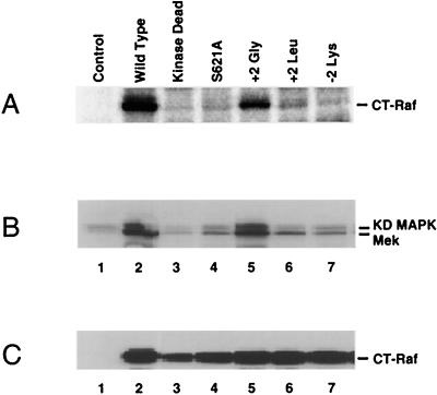
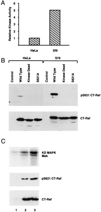
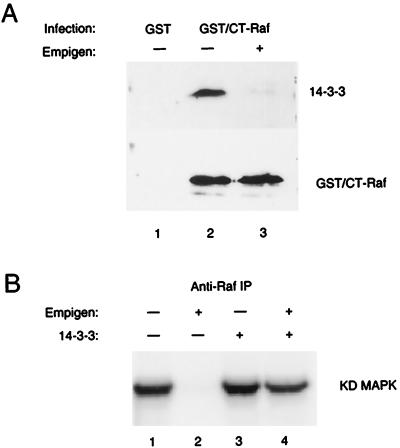
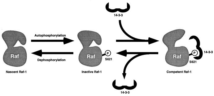
By binding to serine-phosphorylated proteins, 14-3-3 proteins function as effectors of serine phosphorylation. The exact mechanism of their action is, however, still largely unknown. Here we demonstrate a requirement for 14-3-3 for Raf-1 kinase activity and phosphorylation. Expression of dominant negative forms of 14-3-3 resulted in the loss of a critical Raf-1 phosphorylation, while overexpression of 14-3-3 resulted in enhanced phosphorylation of this site. 14-3-3 levels, therefore, regulate the stoichiometry of Raf-1 phosphorylation and its potential activity in the cell. Phosphorylation of Raf-1, however, was insufficient by itself for kinase activity. Removal of 14-3-3 from phosphorylated Raf abrogated kinase activity, whereas addition of 14-3-3 restored it. This supports a paradigm in which the effects of phosphorylation on serine as well as tyrosine residues are mediated by inducible protein-protein interactions.


Figures

FIG. 1
FIG. 1
Binding of mutated 14-3-3η proteins to a serine-phosphorylated Raf peptide. A schematic representation of the helices comprised by a 14-3-3 dimer, with the relative locations of amino acid substitutions introduced into 14-3-3η by site-directed mutagenesis, is shown. Each mutant was expressed as a GST fusion protein, and binding of the proteins at 1 μM to a serine-phosphorylated Raf peptide by SPR was compared at equilibrium. Shown is the average percent binding ± one standard deviation (SD) determined in two separate experiments for each mutated protein relative to the binding obtained with the wild-type 14-3-3η fusion protein at an equal concentration. Helices shown in gray are those expected to contain amino acids lining the proposed binding surface.
FIG. 2
FIG. 2
Binding-impaired 14-3-3 proteins inhibit CT-Raf kinase activity. (A) Activity of CT-Raf coexpressed with wild-type (WT) or mutated 14-3-3η proteins. 293 cells were transfected with a Ras-responsive reporter plasmid (pB4XCAT) and expression plasmids encoding CT-Raf and the indicated 14-3-3η construct. Cell lysates were analyzed 24 h later for CAT activity. Values were normalized to the amount of activity obtained from CT-Raf alone. Results shown are averages and standard deviations from four experiments. (B) Binding-impaired 14-3-3 proteins have no effect on the activity of constitutively active MEK. 293 cells were transfected as for panel A, using the constitutively active MEK instead of CT-Raf. CAT assays were performed 24 h after transfection, and the results are expressed as described for panel A. (C) Immunoblots of lysates from panel A or B. Equal aliquots of each lysate were separated on 10% gels by SDS-PAGE and transferred to nitrocellulose. Immunoblots were then developed with anti-Raf, anti-14-3-3, or antihemagglutinin (anti-HA) antibodies as indicated. Lane 1, untransfected; lane 2, CT-Raf only; lanes 3 to 6, CT-Raf with wild-type 14-3-3η (lane 3) or the R56,60A (lane 4), R132A (lane 5), or Y216F (lane 6) mutant; lane 7, HA-R4F Mek alone; lanes 8 and 9, HA-R4F Mek with wild-type 14-3-3η (lane 8) or R56,60A 14-3-3η (lane 9). (D) Coexpression of binding-impaired 14-3-3 proteins with CT-Raf destabilizes the phosphorylation of CT-Raf at S621. Equal aliquots of lysates from panel A were separated by SDS-PAGE and transferred to nitrocellulose. The blots were developed with the phosphospecific anti-p621 antibody. Lane 1, untransfected; lanes 2 to 5, CT-Raf with wild-type 14-3-3η (lane 2) or the R56,60A (lane 3), R132A (lane 4), or Y216F (lane 5) mutant.
FIG. 3
FIG. 3
Specificity of the anti-Raf phosphoserine 621 antibody. (A) Tryptic peptide analysis of CT-Raf phosphorylated by C-TAK1. CT-Raf was expressed in bacteria as a GST fusion protein. After purification, it was labeled in vitro with purified C-TAK1 and [γ-32P]ATP and then digested with trypsin. Peptides were resolved by HPLC. Radioactivity associated with each fraction was measured by scintillation counting. (B) Manual Edman degradation of tryptic fraction 51. Fraction 51 from panel A was subjected to manual Edman degradation. Bars represent radioactivity released from the membrane. The starting radioactivity associated with the membrane was 376 cpm. (C) Anti-phospho-S621 immunoblotting. CT-Raf or histidine-tagged Cdc25C was either expressed alone (lanes 1 and 3) or coexpressed with C-TAK1 (lanes 2 and 4) in bacteria. Lanes 1 and 2, cell extracts were analyzed by SDS-PAGE, transferred to nitrocellulose, and immunoblotted with antibodies to Raf-1 (top panel) or with anti-p621 (lower panel). Lanes 3 and 4, His-tagged Cdc25C was purified by nickel chromatography and Coomassie blue stained (upper panel) or blotted with anti-p621 (lower panel).
FIG. 4
FIG. 4
Analysis of the effect of amino acid substitutions at the +2 or −2 position relative to pS621 in Raf on 14-3-3 binding. (A) Raf phosphopeptides containing the indicated amino acid substitution at the +2 position relative to S621 were assessed for the ability to inhibit the binding of a 14-3-3η fusion protein to immobilized, wild-type Raf phosphopeptide. For each mutated peptide, the concentration required to achieve 50% inhibition (IC50) of 14-3-3 binding is shown. Essentially identical results were obtained with 14-3-3β, 14-3-3τ, and 14-3-3ζ (data not shown). (B) Raf phosphopeptides containing the indicated amino acid substitution at the −2 position relative to S621 were assessed as for panel A. Shown are the peptide concentrations required to achieve 50% inhibition of 14-3-3 binding to the wild-type peptide. Essentially identical results were obtained with 14-3-3β, 14-3-3τ, and 14-3-3ζ (data not shown).
FIG. 5
FIG. 5
S621 phosphorylation and kinase activity of wild-type or mutated CT-Raf proteins. (A) Phosphate labeling of mutated CT-Raf proteins. Sf9 cells infected with recombinant baculoviruses encoding the indicated CT-Raf constructs or uninfected cells (control) were cultured in the presence of [32P]orthophosphoric acid for 12 h. Cells were lysed in NP-40 lysis buffer, and Raf-1 immunoprecipitates were prepared. After resolution by SDS-PAGE, the labeled proteins were visualized by phosphorimaging. Equivalent expression of each construct was verified separately by immunoblot analysis (data not shown). The data shown are representative of three separate experiments. (B) In vitro kinase activity of wild-type or mutated CT-Raf proteins. Sf9 cells were infected with a recombinant baculovirus encoding MEK-1 alone (control) or coinfected with the MEK-1 virus and a second recombinant baculovirus encoding the indicated CT-Raf construct. At 48 h postinfection, cell lysates were prepared. Anti-MEK-1 immunoprecipitations from each lysate were analyzed for MEK-1 kinase activity by using recombinant, kinase-deficient (KD) MAPK as a substrate. Lane 1, cells infected with MEK-1 alone. Lanes 2 to 7, cells infected with MEK-1 and the following CT-Raf constructs: lane 2, wild type; lane 3, K375M (kinase dead); lane 4, S621A; lane 5, +2 Gly (P623G); lane 6, +2 Leu (P623L); lane 7, −2 Lys (S619K). Data shown are representative of four separate experiments. (C) Expression levels of CT-Raf constructs used for panel B. Equal aliquots of lysates used for panel B were resolved by SDS-PAGE, transferred to nitrocellulose, and developed with an anti-Raf antibody. Lane contents are identical to those in panel B.
FIG. 6
FIG. 6
Kinase activity and S621 phosphorylation of CT-Raf expressed in HeLa or Sf9 cells. (A) Relative in vitro kinase activities of HeLa-expressed CT-Raf and Sf9-expressed CT-Raf. Anti-Raf immunoprecipitates from lysates of HeLa cells expressing FLAG–CT-Raf or Sf9 cells expressing baculovirus-encoded CT-Raf were analyzed for in vitro kinase activity by using a linked assay as described in Materials and Methods. Kinase activity from dried gels was quantitated by using a phosphorimager. CT-Raf protein expression levels from each cell type were quantitated by densitometric analysis of immunoblots prepared with equal aliquots of the lysates. The kinase activity observed from each cell type was then adjusted to reflect equivalent protein levels. (B) Analysis of S621 phosphorylation of CT-Raf constructs expressed in HeLa cells and Sf9 cells. Lysates of HeLa cells or Sf9 cells expressing the indicated CT-Raf construct were adjusted to contain equivalent amounts of CT-Raf protein as judged by anti-Raf immunoblotting (lower panel). Aliquots of each lysate were then immunoblotted with the anti-phospho-S621 antibody (upper panel). (C) Coexpression of 14-3-3 with CT-Raf in HeLa cells augments CT-Raf S621 phosphorylation and kinase activity. HeLa cells were either transfected with CT-Raf alone (lane 2) or CT-Raf plus 14-3-3β (lane 3) or left untransfected (lane 1). Raf immunoprecipitates were prepared and analyzed for in vitro kinase activity by using a linked assay (upper panel) as described in Materials and Methods. Equal aliquots of each lysate were analyzed by immunoblotting with the anti-p621 antibody (middle panel) or an anti-Raf antibody (lower panel). KD, kinase dead.
FIG. 7
FIG. 7
14-3-3 is required for CT-Raf kinase activity. (A) 14-3-3 can be removed from CT-Raf in vitro with the detergent Empigen-BB. Sf9 cells were infected with recombinant baculoviruses encoding either GST or a GST–CT-Raf fusion protein. Forty-eight hours after infection, the GST or GST–CT-Raf proteins were isolated from NP-40 lysates by using glutathione-agarose beads. The bead-bound complexes were then washed with either NP-40 lysis buffer (lanes 1 and 2) or NP-40 lysis buffer containing 1% Empigen-BB (lane 3), resolved by SDS-PAGE, transferred to nitrocellulose, and developed with an antibody to either 14-3-3 (upper panel) or Raf (lower panel). (B) Kinase activity of CT-Raf immunoprecipitates washed with Empigen BB, with or without addition of recombinant 14-3-3. Anti-Raf immunoprecipitates (IP) were prepared from lysates of Sf9 expressing CT-Raf and washed with NP-40 lysis buffer (lanes 2 and 4) or with 1% Empigen-BB (lanes 1 and 3). Following this wash step, purified, recombinant 14-3-3 protein was added as indicated (lanes 3 and 4). The in vitro kinase activity of the immunoprecipitates was then assessed by using a linked assay as described in Materials and Methods. Shown are results from one representative experiment in which recombinant 14-3-3τ was used. Other experiments were performed with recombinant 14-3-3β, with similar results. Quantitation of the 32P-labeled substrate in each lane was performed by volume analysis of the phosphorimaged data. Results for each lane normalized to lane 1: lane 1, 100%; lane 2, 0%; lane 3, 115%; lane 4, 75%. KD, kinase dead.
FIG. 8
FIG. 8
Potential role for 14-3-3 in Raf-1 maturation. 14-3-3 may play a critical role in Raf-1 maturation. Raf-1 molecules may exist in dynamic equilibrium between phosphorylated and unphosphorylated forms. Raf-1 can autophosphorylate itself at S621, but in the absence of 14-3-3, this phosphorylation is rapidly lost in the cell, presumably via the action of a phosphatase. The binding of 14-3-3 to this site protects the phosphorylation from phosphatase activity and is proposed to stabilize a kinase-competent conformation in Raf. This 14-3-3-bound form of CT-Raf possesses constitutive activity, requiring no additional activation events. In the context of the full-length molecule, the binding of 14-3-3 to the pS621 site is proposed to result in a preactivated molecule whose kinase activity is repressed due to interactions with the amino-terminal domains. This form of the kinase would be competent to bind Ras and become activated, or derepressed, at the plasma membrane.

References

    1. Aitken A. 14-3-3 proteins on the MAP. Trends Biochem Sci. 1995;20:95–97. - PubMed
    1. Autieri M V, Haines D S, Romanic A M, Ohlstein E H. Expression of 14-3-3 gamma in injured arteries and growth factor- and cytokine-stimulated human vascular smooth muscle cells. Cell Growth Differ. 1996;7:1453–1460. - PubMed
    1. Avruch J, Zhang X F, Kyriakis J M. Raf meets Ras: completing the framework of a signal transduction pathway. Trends Biochem Sci. 1994;19:279–283. - PubMed
    1. Boston P, Jackson P, Kynoch P, Thompson R. Purification, properties and immunohistochemical localisation of human brain 14-3-3 protein. J Neurochem. 1982;38:1466–1474. - PubMed
    1. Braselmann S, McCormick F. BCR and RAF form a complex in vivo via 14-3-3 proteins. EMBO J. 1995;14:4839–4848. - PMC - PubMed

Publication types

MeSH terms