. 1998 Sep;18(9):5247-55.
doi: 10.1128/MCB.18.9.5247.

Modulated expression of the epidermal growth factor-like homeotic protein dlk influences stromal-cell-pre-B-cell interactions, stromal cell adipogenesis, and pre-B-cell interleukin-7 requirements

Affiliations

Modulated expression of the epidermal growth factor-like homeotic protein dlk influences stromal-cell-pre-B-cell interactions, stromal cell adipogenesis, and pre-B-cell interleukin-7 requirements

S R Bauer et al. Mol Cell Biol. 1998 Sep.

Abstract

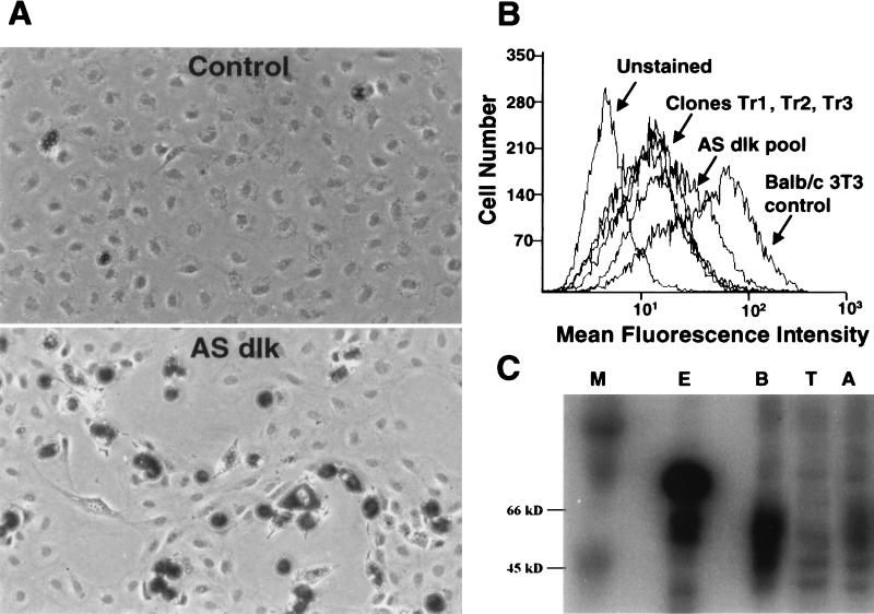
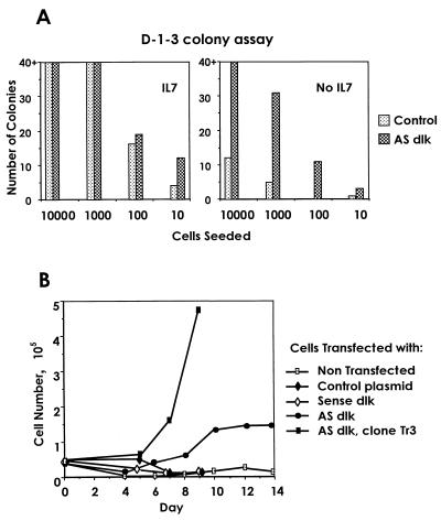
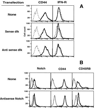
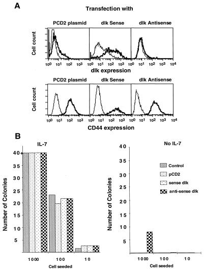
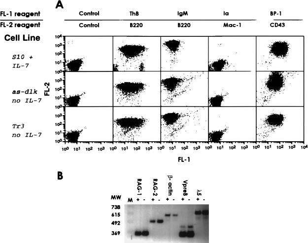
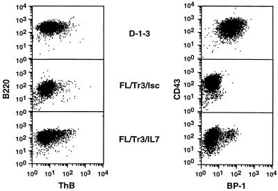
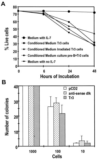
A close relationship exists between adipocyte differentiation of stromal cells and their capacity to support hematopoiesis. The molecular basis for this is unknown. We have studied whether dlk, an epidermal growth factor-like molecule that intervenes in adipogenesis and fetal liver hematopoiesis, affects both stromal cell adipogenesis and B-cell lymphopoiesis in an established pre-B-cell culture system. Pre-B-cell cultures require both soluble interleukin-7 (IL-7) and interactions with stromal cells to promote cell growth and prevent B-cell maturation or apoptosis. We found that BALB/c 3T3 fibroblasts express dlk and function as stromal cells. Transfection of these cells with antisense dlk decreased dlk expression and increased insulin-induced adipocytic differentiation. When antisense transfectants were used as stroma, IL-7 was no longer required to support the growth of pre-B cells and prevent maturation or apoptosis. Antisense dlk transfectants of S10 stromal cells also promoted pre-B-cell growth in the absence of IL-7. These results show that modulation of dlk on stromal cells can influence their adipogenesis and the IL-7 requirements of the pre-B cells growing in contact with them. These results indicate that dlk influences differentiation signals directed both to the stromal cells and to the lymphocyte precursors, suggesting that dlk may play an important role in the bone marrow hematopoietic environment.


Figures

FIG. 1
FIG. 1
Effects of antisense dlk expression on BALB/c 3T3 cells. BALB/c 3T3 cells were transfected with a vector expressing full-length dlk cDNA in antisense orientation. (A) Untransfected (control) cells or pooled antisense transfectants (AS dlk) were treated with 1 μM insulin for 7 to 10 days and then assessed for adipocyte differentiation by Oil-Red O staining as described in Materials and Methods. Dark areas indicate lipid accumulation. (B) Cell surface expression of dlk was examined by flow cytometry by using dlk-specific rabbit antiserum as described in Materials and Methods. Mean fluorescence intensity was plotted against cell numbers of untransfected BALB/c 3T3 controls, pooled antisense dlk transfectants (AS dlk), and three individual antisense transfectant clones (Tr1, Tr2, and Tr3). (C) Western blot analysis of dlk expression in cellular extracts from BALB/c 3T3 cells transfected with antisense dlk expression constructs. Lanes: M, molecular size markers; E, Escherichia coli glutathione S-transferase-dlk fusion protein (73-kDa molecular mass); B, nontransfected BALB/c 3T3 cells; T, clone Tr3; A, pool of antisense-dlk-transfected BALB/c 3T3 cells. The multiple dlk bands result from alternate splicing or differences in glycosylation.
FIG. 2
FIG. 2
Effects of diminished stromal cell dlk expression on pre-B-cell growth. (A) Pre-B-cell colony assay. D-1-3 pre-B cells were seeded into 24-well dishes on irradiated BALB/c 3T3 (control) or dlk antisense transfectants (AS dlk) in the presence (left panel) or absence (right panel) of IL-7. Pre-B cells were seeded in 10-fold dilutions (x axis), and the number of colonies that formed at each pre-B-cell dilution (y axis) was plotted. The greatest number of individually discernible colonies was 40, so bars reaching 40+ indicate wells that were virtually confluent. Bar height represents the mean of duplicate wells. (B) Pre-B-cell growth analysis. Pre-B-cell line D-1-3 was seeded into wells of six-well culture dishes with irradiated stromal cell layers in the absence of IL-7. The mean number of viable pre-B cells in duplicate wells harvested on the indicated days is shown for nontransfected BALB/c 3T3 cells or for BALB/c 3T3 cell lines made by transfection with control plasmid containing no insert or the same plasmid containing full-length dlk cDNA either in the sense (Sense dlk) or antisense (AS dlk) orientation. The control, sense, and antisense lines were pooled following selection. Tr3 is a cloned derivative of the AS dlk line.
FIG. 3
FIG. 3
Flow cytometry analysis of transfected BALB/c 3T3 cells. (A) Cells transfected with sense or antisense dlk expression constructs. Expression levels of CD44 and IFN-γ receptor (IFN-γ) are compared. (B) Cells transfected with antisense Notch constructs. Expression levels of Notch, CD44, and CD45RB are indicated. The thicker lines represent specific staining of the corresponding markers, whereas dotted or thinner lines represent the unstained controls.
FIG. 4
FIG. 4
Effects of enforced downregulation of dlk expression on S10 stromal cells on pre-B-cell growth. (A) S10 cells stably transfected with sense or antisense dlk expression constructs were analyzed for their levels of dlk and CD44 expression. The thicker lines represent specific staining of the corresponding markers, whereas the thinner lines represent the unstained controls. (B) Transfected S10 cells were also used as stroma in cell colony assays using the D-1-3 pre-B-cell line. The bar graph represents the number of colonies that develop in relation to the number of pre-B cells seeded per well. The key shows what construct was transfected into S10 cells.
FIG. 5
FIG. 5
Effects of diminished stromal cell dlk expression on pre-B-cell differentiation. (A) Cell surface marker analysis using the indicated antibodies was done on D-1-3 pre-B cells propagated either under normal culture conditions with S10 stromal cells and IL-7 or without IL-7 on low-dlk-expressing antisense transfectants (as-dlk or Tr3). Each dot plot panel displays fluorescence intensity for PE-labeled antibodies (FL-2; y axis) plotted against fluorescence for FITC-labeled antibodies (FL-1; x axis). (B) Gene expression analysis was done by using reverse transcription-PCR on RNAs isolated from D-1-3 pre-B cells propagated either under normal culture conditions with S10 stromal cells and IL-7 (indicated by +) or without IL-7 on low-dlk-expressing antisense transfectant Tr3 cells (indicated by −). The genes studied are indicated at the top of the panel. A 123-bp ladder (lane M) was included for determination of PCR product size. Lengths in base pairs (MW) of standards are shown on the left.
FIG. 6
FIG. 6
Comparison of cell markers expressed by pre-B-cell lines freshly isolated from mouse fetal liver cells cultured over Tr3 stromal cells in the presence or absence of IL-7 and markers expressed by the pre-B-cell line D-1-3 cultured over S10 cells in the presence of IL-7. Developing colonies of fetal liver cells were expanded over Tr3 cells in the presence or absence of IL-7. Cell type abbreviations: FL, fetal liver origin; Tr3, stromal cell used; Isc, Iscove’s medium–4% FCS; IL-7, IL-7 added to the medium. The expression of cellular markers characteristic of the pre-B phenotype, including B220, ThB, CD43, and BP-1, was analyzed by flow cytometry.
FIG. 7
FIG. 7
Effects of several stromal-cell-conditioned media on pre-B-cell growth. (A) Kinetics of apoptosis of D-1-3 pre-B cells cultured over S10 stromal cells in the presence of the indicated conditioned media or in the presence of normal medium with or without IL-7; (B) cell colony growth assay of D-1-3 pre-B cells cultured over S10 stromal cells in the presence of the indicated conditioned media.

References

    1. Aaronson S A, Todaro G J. Development of 3T3-like lines from Balb-c mouse embryo cultures: transformation susceptibility to SV40. J Cell Physiol. 1968;72:141–148. - PubMed
    1. Artavanis-Tsakonas S, Matsuno K, Fortini M E. Notch signaling. Science. 1995;268:225–232. - PubMed
    1. Bauer S R, Scheuermann R H. Expression of the VpreB/5/pseudo-Ig complex correlates with downregulated RAG-1 expression and V(D)J type recombination: a mechanism for allelic exclusion at the IgH locus. Transgenics. 1993;1:33–45.
    1. Boone C W, Scott R E. Plate-induced tumors of BALB/3T3 cells exhibiting foci of differentiation into pericytes, chondrocytes, and fibroblasts. J Supramol Struct. 1980;14:233–240. - PubMed
    1. Chomczynski P, Sacchi N. Single-step method of RNA isolation by acid guanidinium thiocyanate-phenol-chloroform extraction. Anal Biochem. 1987;162:156–159. - PubMed

MeSH terms

LinkOut - more resources