. 1998 Sep;18(9):5284-90.
doi: 10.1128/MCB.18.9.5284.

Requirement of cyclin E-Cdk2 inhibition in p16(INK4a)-mediated growth suppression

Affiliations

Requirement of cyclin E-Cdk2 inhibition in p16(INK4a)-mediated growth suppression

H Jiang et al. Mol Cell Biol. 1998 Sep.

Abstract

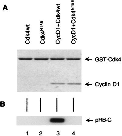
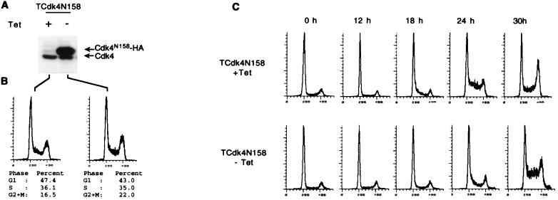
Loss-of-function mutations of p16(INK4a) have been identified in a large number of human tumors. An established biochemical function of p16 is its ability to specifically inhibit cyclin D-dependent kinases in vitro, and this inhibition is believed to be the cause of the p16-mediated G1 cell cycle arrest after reintroduction of p16 into p16-deficient tumor cells. However, a mutant of Cdk4, Cdk4(N158), designed to specifically inhibit cyclin D-dependent kinases through dominant negative interference, was unable to arrest the cell cycle of the same cells (S. van den Heuvel and E. Harlow, Science 262:2050-2054, 1993). In this study, we determined functional differences between p16 and Cdk4(N158). We show that p16 and Cdk4(N158) inhibit the kinase activity of cellular cyclin D1 complexes through different mechanisms. p16 dissociated cyclin D1-Cdk4 complexes with the release of bound p27(KIP1), while Cdk4(N158) formed complexes with cyclin D1 and p27. In cells induced to overexpress p16, a higher portion of cellular p27 formed complexes with cyclin E-Cdk2, and Cdk2-associated kinase activities were correspondingly inhibited. Cells engineered to express moderately elevated levels of cyclin E became resistant to p16-mediated growth suppression. These results demonstrate that inhibition of cyclin D-dependent kinase activity may not be sufficient to cause G1 arrest in actively proliferating tumor cells. Inhibition of cyclin E-dependent kinases is required in p16-mediated growth suppression.


Figures

FIG. 1
FIG. 1
Dominant negative properties of Cdk4N158. (A) Purification of cyclin D1-Cdk4 and cyclin D1-Cdk4N158 complexes from insect cells infected with the indicated recombinant baculoviruses. wt, wild type. The gel was stained with Coomassie blue. (B) In vitro kinase assay on a GST-pRB-C-terminal fragment with the purified Cdk4 or cyclin D1-Cdk4 complexes shown in panel A.
FIG. 2
FIG. 2
U2OS cell lines with inducible expression of p16. (A) Expression of p16 in the presence (+) or absence (−) of tetracycline (Tet) or at different times after the withdrawal of tetracycline was determined by Western blotting of the total cell extracts with an anti-p16 antibody (JC-6). Shown are cell lines Tp16wt-17 and Tp16P114L-11 that are representatives of multiple similar cell lines established in this study. (B) Effects of p16 on cell cycle profiles of asynchronously growing U2OS cells were determined by flow cytometry analysis after propidium iodide staining (15,000 cells were routinely counted for each sample). The scale on the x axes indicates relative propidium iodide staining intensity.
FIG. 3
FIG. 3
U2OS cell lines with inducible expression of Cdk4N158. (A) Expression of Cdk4N158 in the presence (+) or absence (−) of tetracycline (Tet) was determined by Western blotting of the total cell extracts with an anti-Cdk4 antibody (H22). (B) Effects of Cdk4N158 overexpression on cell cycle profiles of asynchronously growing cells were determined by flow cytometry analysis. Scale on x axes indicates relative propidium iodide staining intensity. (C) Cdk4N158 inducible cells were serum starved for 5 days and released into media containing 10% serum in the presence and absence of tetracycline. At the indicated time points after release, an aliquot of cells was harvested for flow cytometry analysis to determine the cell cycle profiles.
FIG. 4
FIG. 4
Effects of p16 and Cdk4N158 on the activities of cellular cyclin-dependent kinases. (A) Kinase activity in the presence (+) or absence (−) of tetracycline (Tet) determined by immunoprecipitation and in vitro phosphorylation assay. Total cell extracts of Tp16wt, Tp16P114L, and TCdk4N158 cells before and 24 h after induction were immunoprecipitated with either anti-cyclin D1 (DCS11) or anti-Cdk2 (M2) antibodies, and kinase reactions were carried out with purified GST-pRB-C-terminal fragment. Reaction products were separated in sodium dodecyl sulfate-polyacrylamide gel electrophoresis (SDS-PAGE) and visualized either by autoradiography or on a StormImager. Quantitation was performed on StormImager with the ImagerQuant software with results obtained from four independent experiments. (B) Histone H1 kinase activity determined by immunoprecipitation of Cdk2 and in vitro kinase assay. Lanes are as marked at the top of panel A. (C) In vivo phosphorylation status of cellular pRB. Total cell extracts from the indicated cells, as marked at the top of panel A, were separated on an SDS–6% PAGE and blotted with anti-pRB monoclonal antibody (XZ77). pRBphos, phosphorylated pRB.
FIG. 5
FIG. 5
Effects of p16 (Tp16wt and Tp16P114L) and Cdk4N158 overexpression on protein levels of cyclin D1 (Cyc D1), cyclin E (Cyc E), cyclin A (Cyc A), Cdk4, Cdk2, and p27 in the presence (+) or absence (−) of tetracycline (Tet). Protein levels were determined by immunoblotting with the indicated antibodies after SDS-PAGE of total cell extracts as indicated. Equal amounts of protein were loaded. In some cases, nonspecific bands are shown as loading controls, and molecular mass markers (in kilodaltons) are included for cyclin E and cyclin A.
FIG. 6
FIG. 6
Effects of p16 (Tp16wt and T16P114L) and Cdk4N158 (TCdk4N158) overexpression in the presence (+) or absence (−) of tetracycline (Tet) on the composition of cellular cyclin-dependent kinases. The status of cyclin D1, Cdk4, Cdk2, and p27 complexes were determined by immunoprecipitation (IP) followed by Western blotting, as indicated. See Materials and Methods for a description of the antibodies used. Cyc, cyclin; αp27IP, anti-p27 IP.
FIG. 7
FIG. 7
Effects of cellular cyclin E (Cyc E) levels on growth suppression activities of p16 and p27. (A) Cyclin E protein levels in U2OS cells and a derivative cyclin E-transfected cell line (UE13) were determined by Western blotting of the total cell extracts with an anti-cyclin E antibody (HE12). (B) Growth suppression activities of p16 and p27 on U2OS and the cyclin E-transfected cell line were determined by transient transfection with pCMVp16 or pCMVp27 together with the transfection marker CD20. Empty vector was used as a baseline control. Transfected cells were analyzed by flow cytometry, and the cell cycle profiles of CD20-positive cells were determined. Differences in G1 percentage cells (Δ G1 %) between vector-transfected cells and cells transfected with p16 or p27 are presented.

References

    1. Alcorta D A, Xiong Y, Phelps D, Hannon G, Beach D, Barrett J C. Involvement of the cyclin-dependent kinase inhibitor p16 (INK4a) in replicative senescence of normal human fibroblasts. Proc Natl Acad Sci USA. 1996;93:13742–13747. - PMC - PubMed
    1. Alevizopoulos K, Vlach J, Hennecke S, Amati B. Cyclin E and c-Myc promote cell proliferation in the presence of p16INK4a and of hypophosphorylated retinoblastoma family proteins. EMBO J. 1997;16:5322–5333. - PMC - PubMed
    1. Baldin V, Lukas J, Marcote M J, Pagano M, Draetta G. Cyclin D1 is a nuclear protein required for cell cycle progression in G1. Genes Dev. 1993;7:812–821. - PubMed
    1. Bates S, Parry D, Bonetta L, Vousden K, Dickson C, Peters G. Absence of cyclin D/cdk complexes in cells lacking functional retinoblastoma. Oncogene. 1994;9:1633–1640. - PubMed
    1. Blain S W, Montalvo E, Massague J. Differential interaction of the cyclin-dependent kinase (Cdk) inhibitor p27Kip1 with cyclin A-Cdk2 and cyclin D2-Cdk4. J Biol Chem. 1996;272:25863–25872. - PubMed

Publication types

MeSH terms