. 1998 Sep;18(9):5380-91.
doi: 10.1128/MCB.18.9.5380.

Dual cyclin-binding domains are required for p107 to function as a kinase inhibitor

Affiliations

Dual cyclin-binding domains are required for p107 to function as a kinase inhibitor

E Castaño et al. Mol Cell Biol. 1998 Sep.

Abstract

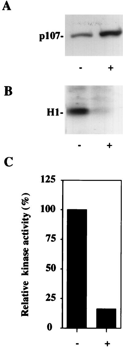
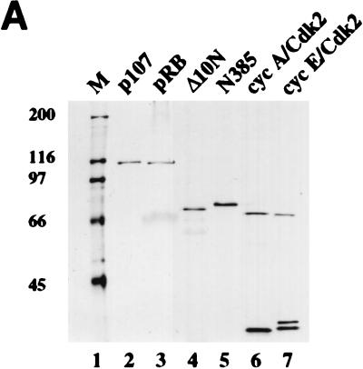
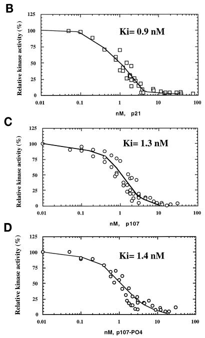
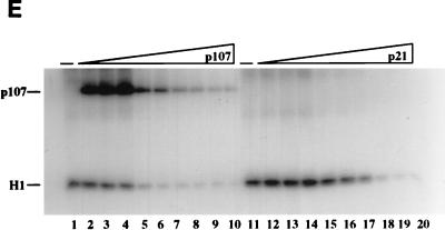
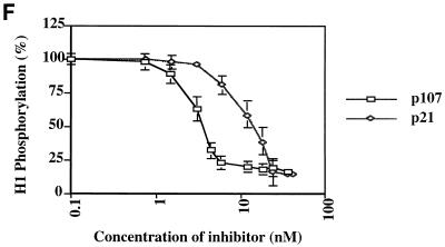
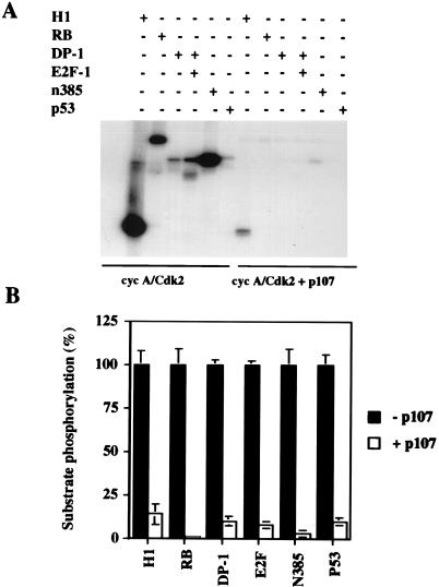
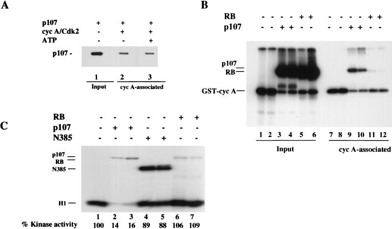
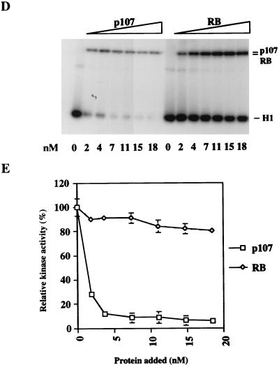
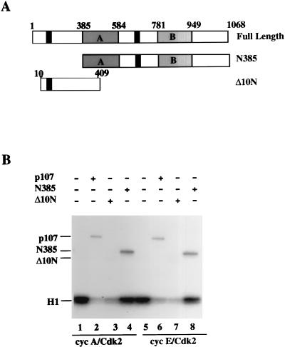
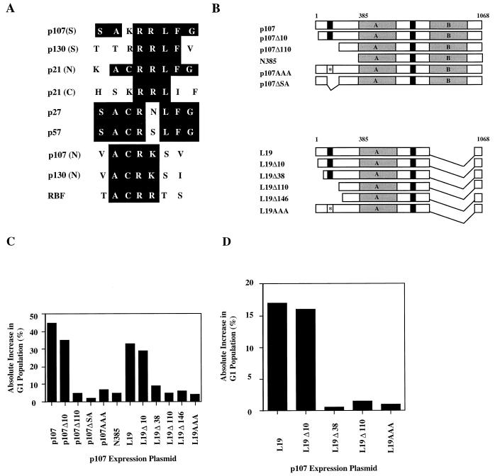
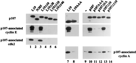
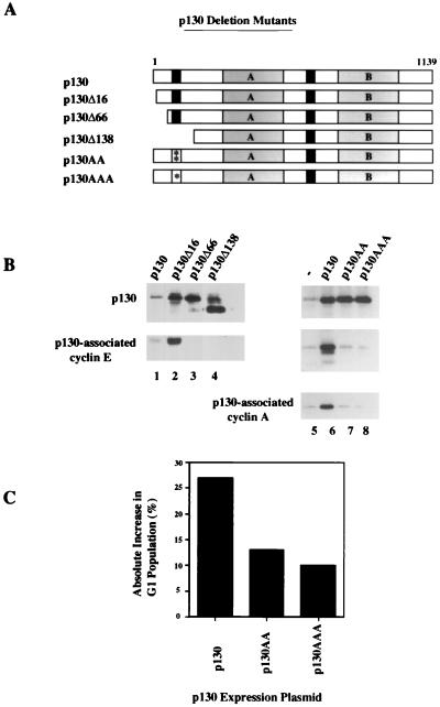
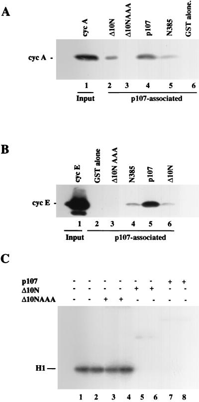
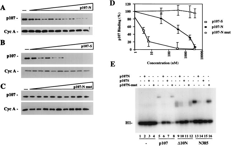
The retinoblastoma (pRB) family of proteins includes three proteins known to suppress growth of mammalian cells. Previously we had found that growth suppression by two of these proteins, p107 and p130, could result from the inhibition of associated cyclin-dependent kinases (cdks). One important unresolved issue, however, is the mechanism through which inhibition occurs. Here we present in vivo and in vitro evidence to suggest that p107 is a bona fide inhibitor of both cyclin A-cdk2 and cyclin E-cdk2 that exhibits an inhibitory constant (Ki) comparable to that of the cdk inhibitor p21/WAF1. In contrast, pRB is unable to inhibit cdks. Further reminiscent of p21, a second cyclin-binding site was mapped to the amino-terminal portions of p107 and p130. This amino-terminal domain is capable of inhibiting cyclin-cdk2 complexes, although it is not a potent substrate for these kinases. In contrast, a carboxy-terminal fragment of p107 that contains the previously identified cyclin-binding domain serves as an excellent kinase substrate although it is unable to inhibit either kinase. Clustered point mutations suggest that the amino-terminal domain is functionally important for cyclin binding and growth suppression. Moreover, peptides spanning the cyclin-binding region are capable of interfering with p107 binding to cyclin-cdk2 complexes and kinase inhibition. Our ability to distinguish between p107 and p130 as inhibitors rather than simple substrates suggests that these proteins may represent true inhibitors of cdks.


Figures

FIG. 1
FIG. 1
p107 and p130 are inhibitors of cdk2 activity in vivo. (A) p107 expression is modestly induced in a tetracycline-repressible cell line by removal of tetracycline. +, p107 induction; −, expression was not induced. (B) Induction of p107 levels shown in panel A causes significant reduction in histone H1 kinase activity. Results of a representative experiment are shown. (C) Quantitation of kinase activity shown in panel B.
FIG. 2
FIG. 2
Inhibition of cyclin A (cyc A)- and cyclin E-cdk2 by p107 and p21 in vitro. (A) Recombinant proteins used in the in vitro assays. p107, pRB, N385, cyclin A-cdk2, and cyclin E-cdk2 were purified from insect cells infected with recombinant baculoviruses. GST-tagged Δ10N was purified from bacteria as described in Materials and Methods. Equal amounts of the indicated proteins were electrophoresed and visualized by silver staining. The sizes of molecular mass markers (lane M) (in kilodaltons) are indicated on the left. (B to D) Histone H1 kinase assays were performed with purified p21 from Escherichia coli or purified p107 (see Materials and Methods). Increasing concentrations of p21 (0.05 to 74 nM), p107 (0.01 to 30 nM), or prephosphorylated p107 (0.01 to 30 nM) were used as indicated. The relative levels of phosphorylated histone H1 were determined with a PhosphorImager. Histone H1 phosphorylation by cyclin A-cdk2 in the absence of an inhibitor was given a value of 100. Calculated average Ki values are indicated. (E) Histone H1 kinase assays comparing p21 and p107 as inhibitors of cyclin E-cdk2. Increasing concentrations of p107 (0.7 to 42 nM) (lanes 2 to 10) or p21 (0.7 to 42 nM) (lanes 12 to 20) were incubated with 1 ng of cyclin E-cdk2. For each set of reactions, a kinase-alone control (lanes −) was included. (F) The relative levels of phosphorylated histone H1 were determined as for panels B to D. Each value represents the mean and standard error of the mean for four independent experiments.
FIG. 2
FIG. 2
Inhibition of cyclin A (cyc A)- and cyclin E-cdk2 by p107 and p21 in vitro. (A) Recombinant proteins used in the in vitro assays. p107, pRB, N385, cyclin A-cdk2, and cyclin E-cdk2 were purified from insect cells infected with recombinant baculoviruses. GST-tagged Δ10N was purified from bacteria as described in Materials and Methods. Equal amounts of the indicated proteins were electrophoresed and visualized by silver staining. The sizes of molecular mass markers (lane M) (in kilodaltons) are indicated on the left. (B to D) Histone H1 kinase assays were performed with purified p21 from Escherichia coli or purified p107 (see Materials and Methods). Increasing concentrations of p21 (0.05 to 74 nM), p107 (0.01 to 30 nM), or prephosphorylated p107 (0.01 to 30 nM) were used as indicated. The relative levels of phosphorylated histone H1 were determined with a PhosphorImager. Histone H1 phosphorylation by cyclin A-cdk2 in the absence of an inhibitor was given a value of 100. Calculated average Ki values are indicated. (E) Histone H1 kinase assays comparing p21 and p107 as inhibitors of cyclin E-cdk2. Increasing concentrations of p107 (0.7 to 42 nM) (lanes 2 to 10) or p21 (0.7 to 42 nM) (lanes 12 to 20) were incubated with 1 ng of cyclin E-cdk2. For each set of reactions, a kinase-alone control (lanes −) was included. (F) The relative levels of phosphorylated histone H1 were determined as for panels B to D. Each value represents the mean and standard error of the mean for four independent experiments.
FIG. 3
FIG. 3
Inhibition of cyclin A (cyc A)-cdk2 substrates. (A) Kinase assays were performed in the absence (left) or presence (right) of 20 ng of p107 (previously phosphorylated). One hundred nanograms of the indicated purified proteins was added to the reaction mixture, with the exception of histone H1, for which 1.25 μg was used. (B) The amount of phosphorylation of each protein was quantified with a PhosphorImager, and the level of phosphorylation of each protein in the absence of p107 was set to 100%. The values represent the means and standard errors of the means for four different experiments.
FIG. 4
FIG. 4
p107 and pRB as inhibitor and substrate, respectively, of cyclin A (cyc A)-cdk2. (A) p107 was allowed to bind cyclin A-cdk2 in the absence or presence of 1 μM ATP for 30 min at 37°C. Glutathione-agarose precipitation of GST-cyclin A-cdk2 was carried out as described in Materials and Methods, followed by immunoblot analysis of p107. The input amount of p107 is shown in lane 1. (B) p107 (lanes 3, 4, 9, and 10) and pRB (lanes 5, 6, 11, and 12) were incubated in a kinase reaction without histone H1 and with [γ-32P]ATP. After 30 min at 37°C, half of the reaction mixture (lanes 1 to 6) was taken and boiled in SDS sample buffer, while the other half (lanes 7 to 12) was incubated with glutathione-agarose beads and precipitated as described in Materials and Methods. (C) Cyclin A-cdk2 kinase reactions in the presence of 20 ng of p107 (lanes 2 and 3), 60 ng of N385 (lanes 4 and 5), or 20 ng of pRB (lanes 6 and 7). (D) An extended titration comparing the effects of equal amounts of pRB and p107 on cyclin A-cdk2 kinase activity. The amount of either protein is indicated below the autoradiogram. (E) Quantitation of the data shown in panel D. The relative percentage of kinase activity was determined in each case by the amount of 32P incorporated into histone H1, as measured with a PhosphorImager.
FIG. 4
FIG. 4
p107 and pRB as inhibitor and substrate, respectively, of cyclin A (cyc A)-cdk2. (A) p107 was allowed to bind cyclin A-cdk2 in the absence or presence of 1 μM ATP for 30 min at 37°C. Glutathione-agarose precipitation of GST-cyclin A-cdk2 was carried out as described in Materials and Methods, followed by immunoblot analysis of p107. The input amount of p107 is shown in lane 1. (B) p107 (lanes 3, 4, 9, and 10) and pRB (lanes 5, 6, 11, and 12) were incubated in a kinase reaction without histone H1 and with [γ-32P]ATP. After 30 min at 37°C, half of the reaction mixture (lanes 1 to 6) was taken and boiled in SDS sample buffer, while the other half (lanes 7 to 12) was incubated with glutathione-agarose beads and precipitated as described in Materials and Methods. (C) Cyclin A-cdk2 kinase reactions in the presence of 20 ng of p107 (lanes 2 and 3), 60 ng of N385 (lanes 4 and 5), or 20 ng of pRB (lanes 6 and 7). (D) An extended titration comparing the effects of equal amounts of pRB and p107 on cyclin A-cdk2 kinase activity. The amount of either protein is indicated below the autoradiogram. (E) Quantitation of the data shown in panel D. The relative percentage of kinase activity was determined in each case by the amount of 32P incorporated into histone H1, as measured with a PhosphorImager.
FIG. 5
FIG. 5
Inhibition of cyclin A (cyc A)- and cyclin E-cdk2 by the amino terminus of p107. (A) Schematic diagram of p107, N385, and Δ10N proteins. Shaded rectangles with A and B denote subdomains of the carboxy-terminal region conserved with pRB and p130, and black rectangles indicate potential amino-terminal and spacer region cyclin-binding domains. (B) Kinase reactions comparing equal amounts (approximately 20 ng) of p107 (lanes 2 and 6), Δ10N (lanes 3 and 7), and N385 (lanes 4 and 8). All of the reaction mixtures contained 1.25 μg of histone H1 as a substrate for cyclin A-cdk2 (lanes 1 to 4) or cyclin E-cdk2 (lanes 5 to 8), and each had been preincubated at room temperature in the presence of unlabeled 1 μM ATP. The positions of phosphorylated histone H1 and p107 proteins are indicated at the left.
FIG. 6
FIG. 6
An amino-terminal region in p107 is important for growth suppression. (A) Schematic alignment of cyclin-binding domains found in the p21-p27-p57 and p107-p130 families of proteins. Previously identified sequences found in the spacer regions (S) of p107 and p130 are indicated. Homologous amino-terminal sequences (N) found in p107, p130, and the Drosophila pRB-related factor, RBF, are shown. (B) Diagram of p107 constructs used in transfection assays. Symbols used are as indicated in the legend to Fig. 5. Deletions and triple alanine point mutations (asterisks) are indicated. (C) Growth arrest of C33A cells mediated by p107 as determined by FACS analysis (see Materials and Methods). The ordinate shows the absolute difference in percentage of cells in the G1 phase of the cell cycle. Values represent averages for at least three independent experiments. (D) Growth arrest of Saos-2 cells by p107 L19 derivatives.
FIG. 7
FIG. 7
Mutations in p107 abolish binding to cyclins A and E. Each of the indicated proteins was expressed in C33A cells by transfection, and whole-cell extracts were immunoprecipitated with antibodies against p107 or the HA tag (which recognize the carboxy-terminal tag on p107 constructs). Immunoprecipitates were electrophoresed and subjected to immunoblotting with the indicated antibodies. A negative control transfection with empty expression vector (lane −) is indicated.
FIG. 8
FIG. 8
Mutations in p130 similar to those introduced into p107 also abolish cyclin binding and diminish growth-suppressive activity. (A) Schematic of p130 mutations. Symbols are described in legend to Fig. 6. (B) Cyclin-binding activity of transfected p130 constructs. Endogenous cyclins were coimmunoprecipitated with p130 by using anti-p130 antibodies as described in the legend to Fig. 7. (C) Growth arrest of C33A cells as determined in Fig. 6C. The transfected p130 plasmids are indicated below the graph, and results represent averages for several independent experiments.
FIG. 9
FIG. 9
Mutation of the p107 amino-terminal cyclin-binding domain abolishes kinase inhibition and binding. Cyclin A (cyc A) (A) and cyclin E (B) bind Δ10N and N385, albeit less efficiently than full-length p107, but do not bind the mutant Δ10N-AAA. (A) Western blot analysis of cyclin A bound to p107 derivatives. Thrombin-cleaved cyclin A, devoid of a GST tag, was tested for binding to GST alone (lane 2), Δ10N (lane 3), Δ10N-AAA (lane 4), p107 (lane 5), and N385 (lane 6). The input amount of cyclin A is shown in lane 1. (B) Western blot analysis of cyclin E bound to p107 derivatives. Thrombin-cleaved cyclin E, devoid of a GST tag, was tested for binding to GST alone (lane 2), Δ10NAAA (lane 3), N385 (lane 4), p107 (lane 5), and Δ10N (lane 6). The cyclin E input is indicated in lane 1. In each case, precipitation of N385 and p107 was performed with a human papillomavirus E7 peptide linked to Sepharose beads, and precipitation of Δ10N and Δ10N-AAA was performed with glutathione-agarose beads as described in Materials and Methods. (C) Kinase reactions comparing equal amounts of Δ10N-AAA (lanes 3 and 4), Δ10N (lanes 5 and 6), or p107 (lanes 7 and 8).
FIG. 10
FIG. 10
Peptide binding competition of p107 to cyclin A (Cyc A)-cdk2. (A to C) Western blot analysis carried out after GST precipitation of GST-cyclin A-cdk2 shows the capacity of peptides p107S and p107N, but not p107N-mut, to compete the binding of p107 to cyclin A-cdk2. The concentrations of peptides range from 2 nM to 5 μM. The amount of cyclin A retained was not affected by addition of peptide and was included as a control. (D) Western blots were densitometrically scanned and quantified by utilizing NIH Image 1.61 software. The values indicate the means and standard errors of the means for three independent experiments. (E) Cyclin A-cdk2 kinase reaction. The peptides p107S and p107N, but not p107N-mut, were able to partially reverse the inhibition of cyclin A-cdk2 by p107 or Δ10N. The peptides had no effect on the kinase reaction by themselves (compare lane 1 to lanes 2 to 4).

References

    1. Adams P D, Sellers W R, Sharma S K, Wu A D, Nalin C M, Kaelin W G. Identification of a cyclin-cdk2 recognition motif present in substrates and p21-like cyclin-dependent kinase inhibitors. Mol Cell Biol. 1996;16:6623–6633. - PMC - PubMed
    1. Beijersbergen R L, Carlee L, Verkhoven R M, Bernards R. Regulation of the retinoblastoma protein-related p107 by G1 cyclin complexes. Genes Dev. 1995;9:1340–1353. - PubMed
    1. Cao L, Faha B, Dembski M, Tsai L-H, Harlow E, Dyson N. Independent binding of the retinoblastoma protein and p107 to the transcription factor E2F. Nature. 1992;355:176–179. - PubMed
    1. Chen I-T, Akamatsu M, Smith M L, Lung F-D T, Duba D, Roller P P, Fornace A J, O’Connor P M. Characterization of p21cip1/waf1 peptide domains required for cyclin E/cdk2 and PCNA interactions. Oncogene. 1996;12:595–607. - PubMed
    1. Chen J, Saha P, Kornbluth S, Dynlacht B D, Dutta A. Cyclin-binding motifs are essential for the function of p21CIP1. Mol Cell Biol. 1996;16:4673–4682. - PMC - PubMed

Publication types

MeSH terms

LinkOut - more resources