. 1998 Sep;18(9):5523-32.
doi: 10.1128/MCB.18.9.5523.

The ability of CD40L, but not lipopolysaccharide, to initiate immunoglobulin switching to immunoglobulin G1 is explained by differential induction of NF-kappaB/Rel proteins

Affiliations

The ability of CD40L, but not lipopolysaccharide, to initiate immunoglobulin switching to immunoglobulin G1 is explained by differential induction of NF-kappaB/Rel proteins

S C Lin et al. Mol Cell Biol. 1998 Sep.

Abstract

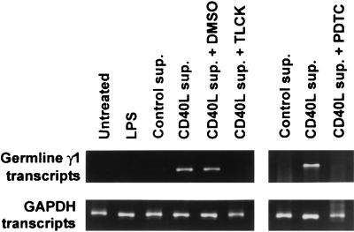
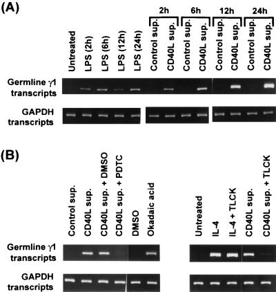
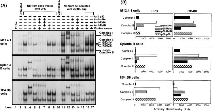
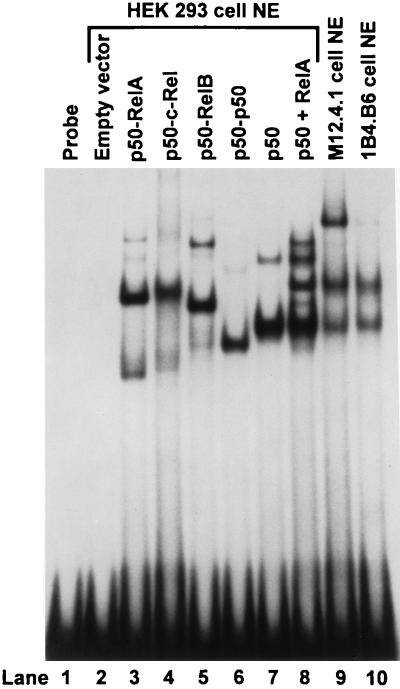
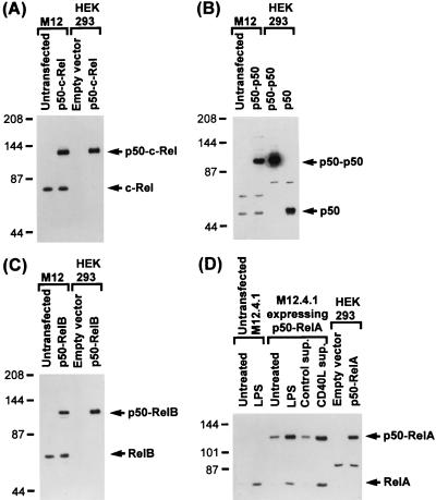
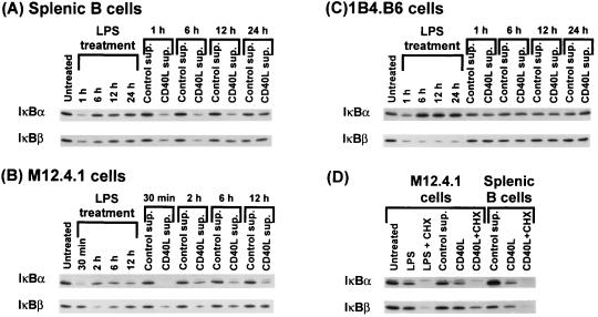
Antibodies of the immunoglobulin G1 class are induced in mice by T-cell-dependent antigens but not by lipopolysaccharide (LPS). CD40 engagement contributes to this preferential isotype production by activating NF-kappaB/Rel to induce germ line gamma1 transcripts, which are essential for class switch recombination. Although LPS also activates NF-kappaB, it poorly induces germ line gamma1 transcripts. Western blot analyses show that CD40 ligand (CD40L) induces all NF-kappaB/Rel proteins, whereas LPS activates predominantly p50 and c-Rel. Electrophoretic mobility shift assays show that in CD40L-treated cells, p50-RelA and p50-RelB dimers are the major NF-kappaB complexes binding to the germ line gamma1 promoter, whereas in LPS-treated cells, p50-c-Rel and p50-p50 dimers are the major binding complexes. Transfection of expression plasmids for NF-kappaB/Rel fusion proteins (forced dimers) indicates that p50-RelA and p50-RelB dimers activate the germ line gamma1 promoter and that p50-c-Rel and p50-p50 dimers inhibit this activation by competitively binding to the promoter without activating the promoter. Therefore, germ line gamma1 transcription depends on the composition of NF-kappaB/Rel proteins.


Figures

FIG. 1
FIG. 1
RT-PCR analysis to demonstrate induction of GL γ1 transcripts in splenic B cells. Resting splenic B cells were left untreated or were treated with LPS (50 μg/ml), control supernatant (sup.) (20%), or CD40L supernatant (20%) for 6 h in the presence or absence of the NF-κB inhibitor PDTC (50 μM) or TLCK (75 μM). Expression of GL γ1 transcripts was assayed by RT-PCR with amplification of GAPDH transcripts as the internal control.
FIG. 2
FIG. 2
RT-PCR analysis of induction of GL γ1 transcripts in 1B4.B6 B cells by LPS, CD40L, and okadaic acid. (A) 1B4.B6 cells were left untreated or were treated with LPS, control supernatant (sup.), or CD40L supernatant, and cells were harvested at the time points indicated for detection of GL γ1 transcripts by RT-PCR. (B) Expression of GL γ1 transcripts was analyzed in 1B4.B6 cells, untreated or treated with control supernatant CD40L supernatant, or IL-4 (1,000 U/ml) for 6 h in the presence or absence of the NF-κB inhibitor PDTC or TLCK. Induction of transcripts by okadaic acid (125 ng/ml), an NF-κB activator, was evaluated at 6 h.
FIG. 3
FIG. 3
Western blot analyses of NF-κB activation by CD40L and LPS treatment. Nuclear extracts from splenic B cells (A) and from the mouse B-cell lines M12.4.1 (B) and 1B4.B6 (C), left untreated or treated with LPS, control supernatant (sup.), or CD40L supernatant for the indicated times, were analyzed. (D) Western blot analysis with nuclear extracts from M12.4.1 B cells untreated or treated for 6 h with control supernatant, CD40L supernatant, or LPS in the presence or absence of CHX (5 μg/ml). Each Western blot was probed sequentially with four antibodies against different NF-κB/Rel proteins. These experiments were performed two or three times with nearly identical results.
FIG. 4
FIG. 4
EMSA and densitometry analyses to compare NF-κB activation by LPS and CD40L. (A) Labeled CD40RR probe was incubated with nuclear extracts (NE) from M12.4.1, 1B4.B6, and splenic B cells treated with CD40L or LPS for 12 h to determine binding activity of each NF-κB/Rel dimer. The three major DNA-protein complexes formed are indicated. To supershift or deplete the specific NF-κB/Rel dimers, nuclear extracts were preincubated with antibody against a specific NF-κB/Rel protein, a control serum, or the indicated combinations of antibodies before incubation with labeled CD40RR probe. The total amount of antibody was kept constant at 3 μl by adding control antibody. (B) Densitometry analyses of the NF-κB/Rel complexes from EMSA results shown in panel A. The quantity of binding activity of complex I, containing p50-RelB, was obtained by subtracting the signals at the position of complex I in lanes 8 and 16 from the signals of complex I in lanes 3 and 11, respectively. The quantity of complex III, containing p50-p50, was obtained by subtracting the signals at the position of complex III in lanes 4 and 12 from the signals of complex III in lanes 3 and 11, respectively. The quantity of p50–c-Rel in complex II was obtained by subtracting the signals of complex II in lanes 5 and 13 from the signals of complex II in lanes 3 and 11, respectively. The quantity of p50-RelA in complex II was obtained by subtracting the signals of complex II in lanes 6 and 14 from the signals of complex II in lanes 5 and 13, respectively. The quantity of p50-RelB in complex II was obtained by subtracting the signals at the position of complex II in lanes 7 and 15 from the signals of complex II in lanes 6 and 14, respectively.
FIG. 5
FIG. 5
EMSA to demonstrate the ability of NF-κB/Rel fusion proteins to bind the CD40RR. EMSA was performed with labeled CD40RR fragment as the probe and 2 μg of nuclear extracts (NE) from HEK 293 cells transiently transfected with empty vector or with expression plasmids for p50-RelA, p50–c-Rel, p50-RelB, and p50-p50 fusion proteins (lanes 2 to 6). Nuclear extracts from cells transfected with p50 plasmid alone (lane 7) or both p50 and RelA plasmids (lane 8) demonstrate formation of multiple NF-κB complexes when two NF-κB/Rel subunits are coexpressed. Nuclear extracts from M12.4.1 cells (3 μg) (lane 9) and 1B4.B6 cells (6 μg) (lane 10) treated with CD40L for 12 h illustrate complexes formed by endogenous NF-κB/Rel dimers.
FIG. 6
FIG. 6
Western blot analyses demonstrate that NF-κB/Rel fusion proteins can be stably expressed and translocated into the nucleus in M12.4.1 and HEK 293 cells. (A to C) Nuclear extracts were prepared from M12.4.1 cells (stably) or HEK 293 cells (transiently) transfected with the p50–c-Rel, p50-p50, or p50-RelB plasmid. The fusion proteins and endogenous NF-κB proteins in the nuclear extracts were detected by Western blot analyses using anti-c-Rel, anti-p50, and anti-RelB antibodies. (D) M12.4.1 cells, untransfected or stably transfected with the p50-RelA expression plasmid, were left untreated or were treated with LPS, control supernatant (sup.), or CD40L supernatant for 6 h. p50-RelA and RelA in the nuclear extracts were detected by using anti-RelA in the Western blot analysis. Expression of intact p50-RelA in transiently transfected HEK 293 cells was also detected in the nucleus.
FIG. 7
FIG. 7
Reporter gene assays demonstrate that overexpression of p50–c-Rel or p50-p50 fusion protein suppresses the transactivation activity of p50-RelA and p50-RelB fusion proteins. The −954WT luciferase reporter plasmid (20 μg) that contains the GL γ1 promoter and plasmid pFosCat (15 μg) as the transfection control were cotransfected into 2 × 10⁷ M12.4.1 cells with empty vector or different amounts of expression plasmids for NF-κB/Rel subunits or fusion proteins, as indicated (in picomoles). Empty vector was added to equalize the amount of DNA in each transfection. Luciferase activity, representing the promoter activity, was assayed 9 h after transfection and normalized by CAT activity. The fold induction was calculated by the ratio of the luciferase activity from cells transfected with NF-κB/Rel plasmids to the luciferase activity from cells transfected with empty vector. The mean and standard error (SE) of fold induction were calculated from at least three independent experiments. Similar experiments performed with the ɛ-162Luc plasmid containing the mouse GL ɛ promoter fragment as the reporter plasmid demonstrate that p50–c-Rel is able to transactivate the mouse GL ɛ promoter.
FIG. 8
FIG. 8
Western blot analyses demonstrate induction of IκB degradation by CD40L and LPS. Cytoplasmic extracts (15 μg) from splenic B (A), M12.4.1 (B), and 1B4.B6 (C) cells, left untreated or treated with LPS, control supernatant (sup.), or CD40L supernatant for the indicated times, were analyzed by Western blotting. (D) Cytoplasmic extracts from M12.4.1 and splenic B cells, left untreated or treated for 6 h with control supernatant, CD40L supernatant, or LPS in the presence or absence of CHX, were used in Western blot analyses. Each Western blot was analyzed with antibodies against IκBα and IκBβ proteins sequentially.

References

    1. Baldwin A S., Jr The NF-κB and I-κB proteins: new discoveries and insights. Annu Rev Immunol. 1996;14:649–683. - PubMed
    1. Beg A A, Sha W C, Bronson R T, Ghosh S, Baltimore D. Embryonic lethality and liver degeneration in mice lacking the RelA component of NF-κB. Nature. 1995;376:167–170. - PubMed
    1. Berton M T, Linehan L A. IL-4 activates a latent DNA-binding factor that binds a shared IFN-γ and IL-4 response element present in the germ-line γ1 Ig promoter. J Immunol. 1995;154:4513–4525. - PubMed
    1. Bottaro A, Lansford R, Xu L, Zhang J, Rothman P, Alt F. S region transcription (per se) promotes basal IgE class switch recombination but additional factors regulate the efficiency of the process. EMBO J. 1994;13:665–674. - PMC - PubMed
    1. Brasier A, Tate J, Habener J. Optimized use of the firefly luciferase assay as a reporter gene in mammalian cell lines. BioTechniques. 1989;7:1116–1122. - PubMed

Publication types

MeSH terms