. 1998 Sep;18(9):5609-19.
doi: 10.1128/MCB.18.9.5609.

Fos family members induce cell cycle entry by activating cyclin D1

Affiliations

Fos family members induce cell cycle entry by activating cyclin D1

J R Brown et al. Mol Cell Biol. 1998 Sep.

Abstract

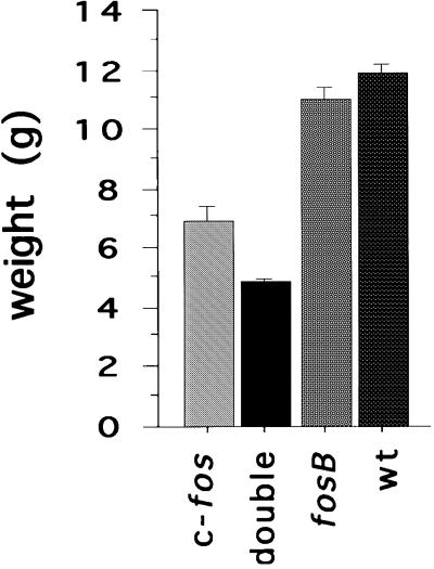
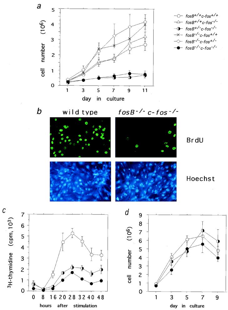
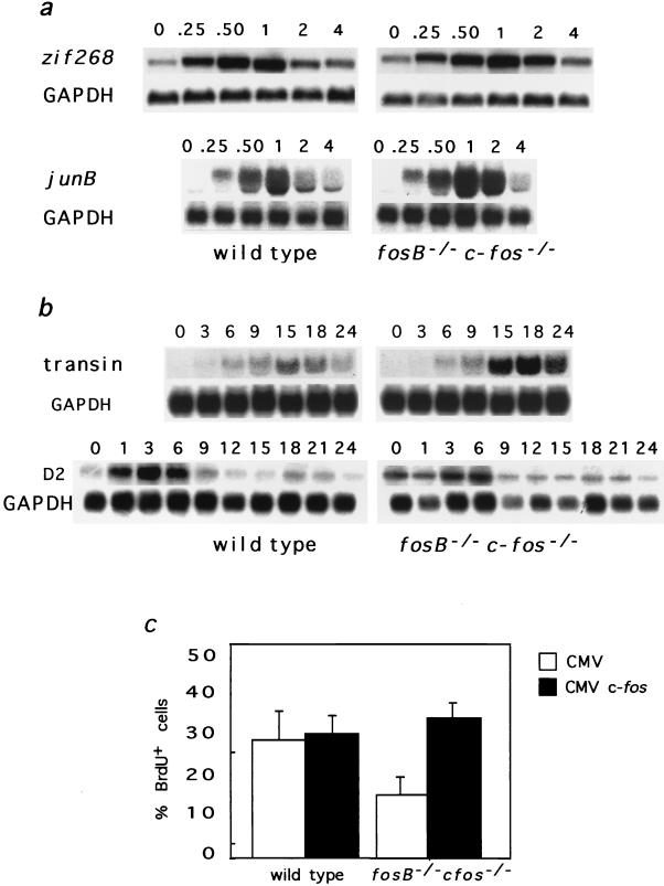
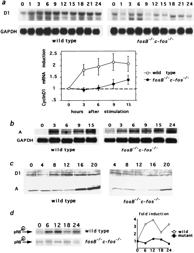
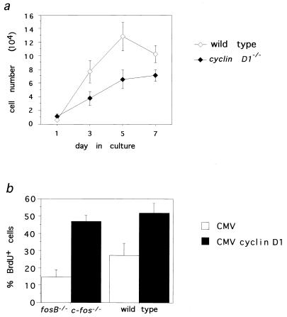
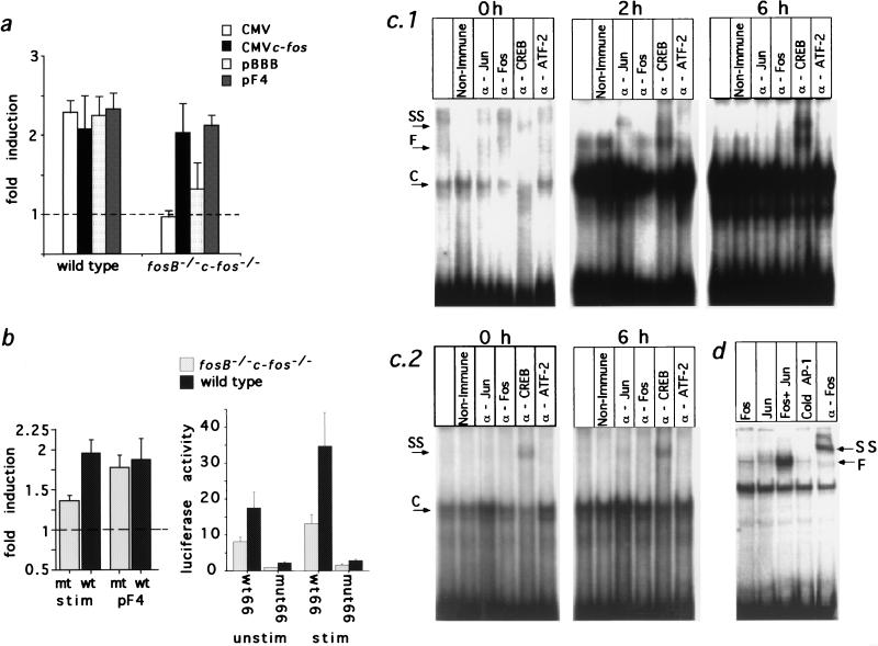
Expression of the fos family of transcription factors is stimulated by growth factors that induce quiescent cells to reenter the cell cycle, but the cellular targets of the Fos family that regulate cell cycle reentry have not been identified. To address this issue, mice that lack two members of the fos family, c-fos and fosB, were derived. The fosB-/- c-fos-/- mice are similar in phenotype to c-fos-/- mice but are 30% smaller. This decrease in size is consistent with an abnormality in cell proliferation. Fibroblasts derived from fosB-/- c-fos-/- mice were found to have a defect in proliferation that results at least in part from a failure to induce cyclin D1 following serum-stimulated cell cycle reentry. Although definitive evidence that c-Fos and FosB directly induce cyclin D1 transcription will require further analysis, these findings raise the possibility that c-Fos and FosB are either direct or indirect transcriptional regulators of the cyclin D1 gene and may function as a critical link between serum stimulation and cell cycle progression.


Figures

FIG. 1
FIG. 1
Average weights of fosB−/− c-fos−/− mice (double) and c-fos−/− mice (c-fos) compared to fosB−/− mice (fosB) and wild-type mice (wt). The weight of each animal in five litters containing fosB−/− c-fos−/− mice was determined between 19 and 23 days of age and combined with previous data on wild-type and fosB−/− mice at the same ages. The difference in weight between fosB−/− c-fos−/− mice and c-fos−/− mice is statistically significant (P < 0.03). Between wild-type or fosB−/− mice and fosB−/− c-fos−/− mice or c-fos−/− mice, all P values are <0.0001.
FIG. 2
FIG. 2
(a) Fibroblast growth at low density. On the indicated day after plating, two plates were harvested and counted. fosB−/− c-fos−/− versus fosB+/− c-fos−/−, no significant difference by repeated-measures analysis of variance (see Materials and Methods). Between the four genotypes that grow well and the two genotypes that grow poorly, all P values are <0.0001. (b) BrdU incorporation following 20 h of stimulation (upper panels) and Hoechst staining of the same fields (lower panels). Magnification, ca. ×33. (c) Incorporation of [3H]thymidine into DNA following serum starvation and stimulation for the indicated number of hours. fosB+/+ c-fos+/+ versus fosB+/− c-fos−/−, P = 0.001; fosB+/+ c-fos+/+ versus fosB−/− c-fos−/−, P = 0.0003; fosB+/− c-fos−/− versus fosB−/− c-fos−/−, no significant difference. (d) Fibroblast growth at high density. No significant differences in cell number were found.
FIG. 3
FIG. 3
(a and b) Northern blots. The upper panels include the gene of interest, and the lower panels include glyceraldehyde-3-phosphate dehydrogenase (GAPDH). Each lane contains 10 μg of total RNA, and all times are hours of stimulation. Shown are two representative IEGs, zif268 and junB (a), and two late genes, cyclin D2 and transin (b). (c) Rescue of S-phase entry by expression of c-fos in continuous cycling conditions. White bars, vector-transfected fibroblasts; black bars, c-fos-transfected fibroblasts. Vector-transfected versus c-fos-transfected fosB−/− c-fos−/− fibroblasts, P = 0.006.
FIG. 4
FIG. 4
(a) Northern blot of cyclin D1 mRNA following serum starvation and stimulation for the indicated number of hours, with glyceraldehyde-3-phosphate dehydrogenase (GAPDH) control. The graphed values are means ± standard errors of cyclin D1 values normalized to GAPDH values for four Northern blots quantitated on a phosphorimager. fosB+/+ c-fos+/+ versus fosB−/− c-fos−/−, P < 0.04. (b) Cyclin A mRNA induction following serum starvation and stimulation, with GAPDH control. (c) Western blots of cyclins D1 (top) and A (bottom) following serum starvation and stimulation for the indicated number of hours. (d) Cyclin D1-associated cdk activity, with glutathione S-transferase–RB as substrate. Immune-complex kinase assays were performed on lysates from wild-type and fosB−/− c-fos−/− fibroblasts following serum starvation and stimulation for the indicated number of hours. The graphed values represent quantitation of the experimental data shown in the figure, which is representative of three performed; in this case, the wild type is actually fosB+/− c-fos+/+. Horizontal axis, number of hours of stimulation.
FIG. 5
FIG. 5
(a) Growth curve of cyclin D1−/− fibroblasts cultured at densities similar to those in Fig. 2a. Cell numbers were determined on the indicated days. Each point for cyclin D1−/− fibroblasts represents the mean value ± standard error of four experiments conducted at least in duplicate. Each point for wild-type fibroblasts represents the mean value ± standard error of at least two experiments conducted in duplicate. Cyclin D1+/+ versus cyclin D1−/−, P < 0.03. (b) Rescue of S-phase entry by expression of cyclin D1 in fosB−/− c-fos−/− fibroblasts in continuous cycling conditions. The ordinate shows the percentage of transfected cells that had incorporated BrdU. CMV indicates vector-transfected fibroblasts, and CMV cyclin D1 indicates cyclin D1-transfected fibroblasts. P is 0.008 for the difference between vector-transfected and cyclin D1-transfected fosB−/− c-fos−/− fibroblasts, and P is 0.0002 for the same difference in wild-type fibroblasts.
FIG. 6
FIG. 6
(a) Induction of the cyclin D1 promoter. Wild-type and fosB−/− c-fos−/− fibroblasts were cotransfected with a cyclin D1-luciferase reporter construct (−1745CD1LUC) and an empty vector control (CMV or pBBB) or a c-fos-containing expression vector (CMV c-fos or pF4). Fold induction is the mean ± standard error of luciferase activity in serum-stimulated samples normalized to the mean ± standard error of luciferase activity in serum-starved samples, for two experiments repeated in triplicate. (b) (Left) Induction of −66CD1LUC. Wild-type (wt) and fosB−/− c-fos−/− (mt) fibroblasts were cotransfected with a cyclin D1-luciferase reporter construct (−66CD1LUC) and an empty vector control (CMV or pBBB) or a c-fos-containing expression vector (pF4). “stim” represents luciferase activity of serum-stimulated samples divided by luciferase activity of serum-starved samples, all cotransfected with CMV or pBBB; P is 0.003 for the difference between wild-type and fosB−/− c-fos−/− fibroblasts. pF4 represents luciferase activity of serum-stimulated samples cotransfected with pF4 divided by luciferase activity of serum-starved samples; no significant difference between the wild type and the mutant was found. (Right) Effect of mutation of the CRE/ATF site of −66CD1LUC on luciferase activity in serum-starved (unstim) and serum-stimulated (stim) wild-type and fosB−/− c-fos−/− fibroblasts. wt66 is −66CD1LUC with a wild-type CRE/ATF site, and mut66 is −66CD1LUC with three point mutations in the CRE/ATF site. The ordinate shows the absolute value of luciferase activity. P is <0.0001 for the difference between the wt66 and mut66 constructs; P is 0.001 for the difference between wild-type and fosB−/− c-fos−/− fibroblasts. (c) Gel mobility shift analyses with the CRE/ATF element of the cyclin D1 promoter and lysates from fibroblasts serum starved and stimulated for the indicated times. Lysates were incubated with either nonimmune serum or antibodies to c-Jun, c-Fos, or CREB/CREM. F, c-Fos-containing complexes; C, CREB/CREM-containing complexes; SS, supershifted complexes. 1, wild type; 2, fosB−/− c-fos−/−. (d) Gel mobility shift analysis with in vitro-transcribed and -translated c-Fos and c-Jun. F and SS are as defined above.

References

    1. Aktas H, Cai H, Cooper G M. Ras links growth factor signaling to the cell cycle machinery via regulation of cyclin D1 and the Cdk inhibitor p27KIP1. Mol Cell Biol. 1997;17:3850–3857. - PMC - PubMed
    1. Albanese C, Johnson J, Watanabe G, Eklund N, Vu D, Arnold A, Pestell R G. Transforming p21ras mutants and c-Ets-2 activate the cyclin D1 promoter through distinguishable regions. J Biol Chem. 1995;270:23589–23597. - PubMed
    1. Brown J R, Ye H, Bronson R T, Dikkes P, Greenberg M E. A defect in nurturing in mice lacking the immediate early gene fosB. Cell. 1996;86:297–309. - PubMed
    1. Bruesselbach S, Moehle-Steinlein U, Wang Z-Q, Schreiber M, Lucibello F C, Mueller R, Wagner E F. Cell proliferation and cell cycle progression are not impaired in fibroblasts and ES cells lacking c-Fos. Oncogene. 1995;10:79–86. - PubMed
    1. Busch S J, Sassone-Corsi P. Dimers, leucine zippers and DNA-binding domains. Trends Genet. 1990;6:36–40. - PubMed

Publication types

MeSH terms

LinkOut - more resources