. 1998 Sep 1;18(17):6790-802.
doi: 10.1523/JNEUROSCI.18-17-06790.1998.

Dendrodendritic inhibition in the olfactory bulb is driven by NMDA receptors

Affiliations

Dendrodendritic inhibition in the olfactory bulb is driven by NMDA receptors

N E Schoppa et al. J Neurosci. .

Abstract

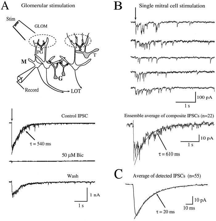
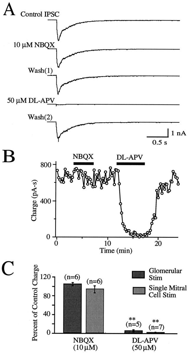
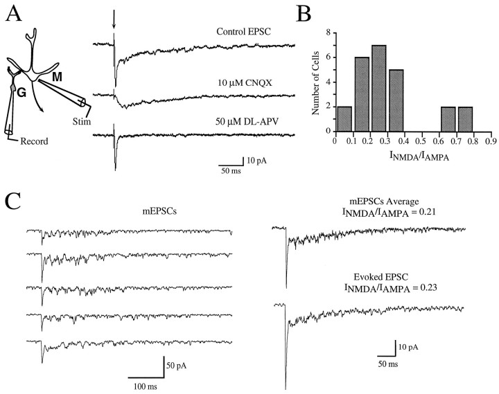
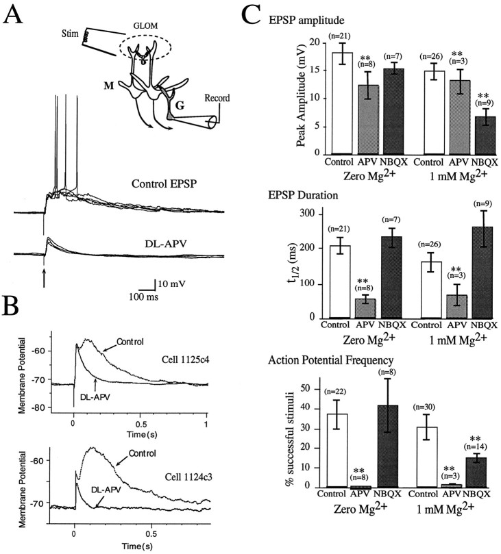
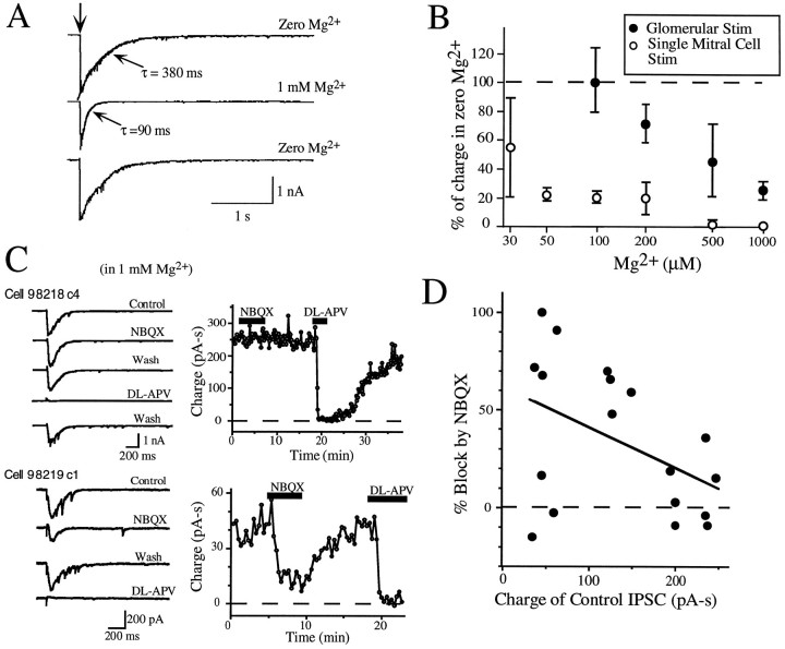
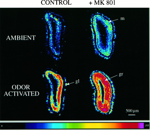
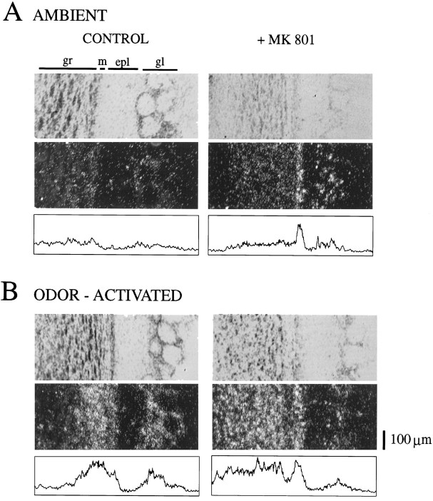
At many central excitatory synapses, AMPA receptors relay the electrical signal, whereas activation of NMDA receptors is conditional and serves a modulatory function. We show here quite a different role for NMDA receptors at dendrodendritic synapses between mitral and granule cells in the rat olfactory bulb. In whole-cell patch-clamp recordings in bulb slices, stimulation of mitral cells elicited slowly decaying, GABAA receptor-mediated reciprocal IPSCs that reflected prolonged GABA release from granule cells. Although granule cells had a normal complement of AMPA and NMDA receptors, the IPSC was completely blocked by the NMDA receptor antagonist D,L-AP-5, suggesting that NMDA receptor activation is an absolute requirement for dendrodendritic inhibition. The AMPA receptor antagonist 1,2,3,4-tetrahydro-6-nitro-2, 3-dioxobenzo[f]quinoxaline-7-sulfonamide (NBQX) had no effect on IPSCs in the absence of extracellular magnesium but modestly reduced IPSCs in 1 mM magnesium, indicating that the primary effect of the AMPA receptor-mediated depolarization was to facilitate the unblocking of NMDA receptors. Granule cell voltage recordings indicated that effective spike stimulation in granule cells depended on the slow NMDA receptor kinetics. Granule cells also showed a pronounced delay between synaptic stimulation and action potential generation, suggesting that their intrinsic membrane properties underlie the ineffectiveness of brief AMPA receptor-mediated EPSPs. NMDA receptors also seem to have a central role in dendrodendritic inhibition in vivo, because intraperitoneal dizocilpine maleate (MK-801) injection in young adult rats resulted in disinhibition of mitral cells as measured by the generation of c-fos mRNA. The unique dependence of dendrodendritic inhibition on slow EPSPs generated by NMDA receptors suggests that olfactory information processing depends on long-lasting reciprocal and lateral inhibition.


Figures

Fig. 1.
Fig. 1.
The kinetics of the dendrodendritic inhibition are determined by the asynchronous release of GABA from granule cells.A, Bipolar stimulation (Stim) in the glomerular layer (GLOM) elicited a long-lasting inward current in the mitral cell (M), when recordings were made with a NaCl extracellular solution with no added magnesium (Control). The current was blocked by bicuculline (Bic; 50 μm), indicating that it reflects a GABAA receptor-mediated IPSC caused by the release of GABA from granule cells (G) at reciprocal dendrodendritic synapses (small circle). Data are from cell 971126c2. In the diagram, individual glomeruli are indicated bydashed ovals; PG, periglomerular cells;T, tufted cell; LOT, lateral olfactory tract. B, Direct somatic stimulation of a mitral cell with the recording pipette also elicited an IPSC, shown here as five raw data traces (top). Thetraces showed unitary IPSCs with fast kinetics, but the averaged, composite IPSC (bottom) had a slow decay (τ = 0.61 sec) like that of the IPSC elicited by glomerular stimulation. The action potential-mediated sodium current was deleted from thesetraces for clarity. Data are from cell 97508c4.C, The average of 55 unitary IPSCs in the recording inB had a decay time constant of 20 msec. Detection of unitary IPSCs was done with a template comprising rising and decaying exponentials (τ of 3 and 30 msec, respectively).
Fig. 2.
Fig. 2.
Dendrodendritic inhibition requires the activation of NMDA receptors. A, The glomerular stimulation-evoked IPSC was insensitive to the AMPA receptor antagonist NBQX (10 μm) but was completely blocked by the NMDA receptor antagonist d,l-AP-5 (50 μm). Each displayedtrace reflects an average of 10–20traces. Data are from cell 971016c5. B, The effect of d,l-AP-5 on the IPSCs in Aoccurred rapidly and was reversible within 5 min after the removal of the drug. Quantification of drug effects was done by integrating the IPSC, yielding a charge value. C, The histogram summarizes the effects of NBQX and d,l-AP-5 on the IPSCs. Similar glutamate receptor pharmacological profiles were observed for the IPSCs evoked by glomerular stimulation and by single mitral cell stimulation.
Fig. 3.
Fig. 3.
Granule cells have both AMPA and NMDA receptors at dendrodendritic synapses. A, Focal mitral cell (M) stimulation (Stim) evoked EPSCs in a granule cell (G) with a CNQX-sensitive, rapidly decaying AMPA receptor-mediated component, as well as a d,l-AP-5-sensitive, slowly decaying NMDA component. The extracellular solution contained no added magnesium. Data are from cell 97617c4. B, The amplitude of the NMDA component was, on average, approximately one-fourth that of the AMPA component. In 24 cells, the ratio of the NMDA and AMPA receptor-mediated currents (INMDA/IAMPA) was taken from the peak current (IAMPA) and the mean current 20–25 msec after stimulus (INMDA).C, mEPSCs in granule cells were recorded in TTX (1 μm) and cadmium (10 μm). Five examples from one cell are shown (left) that were detected using a template that approximated an AMPA mEPSC: a sum of two exponentials with rising and decay components (τ of 0.3 and 3 msec, respectively). The averaged mEPSCs (top right; n = 15) had the same time course as the evoked EPSC in the same cell (bottom right), implying that there are few pure NMDA synapses (see Results). Data are from cell 97812c3.
Fig. 4.
Fig. 4.
Stimulus-evoked action potentials in granule cells require NMDA receptor activation. A, Glomerular (top; GLOM) stimulation (Stim) evoked large dual-component EPSPs in granule cells (G), as well as action potentials. Action potentials were abolished by d,l-AP-5 (bottom; 50 μm). Five responses before and after application of d,l-AP-5 for the same cell are shown superimposed. Firing under control conditions followed a delay, averaging 61 msec in this cell in 13 trials. The bath had no added extracellular magnesium. Data are from cell 1125c4. In the diagram, thedashed oval delineates one glomerulus; M, mitral cell. B, For the cell in A, the peak of the EPSP in d,l-AP-5 (top) was similar to that of the dual-component EPSP; however, the duration, expressed as the time t½ for 50% decay of the voltage signal from the peak, was reduced from 195 to 51 msec. In a different cell (bottom), d,l-AP-5 reduced the amplitude and duration of the EPSP. Only voltage responses that did not elicit action potentials were selected for averaging.C, In the absence of extracellular magnesium,d,l-AP-5 had variable effects on the EPSP amplitude (top) but consistently reduced the durationt½ of the EPSP (middle) and the action potential-firing frequency (bottom). NBQX had little effect on the granule cell voltage responses in no magnesium but had modest effects on the EPSP amplitude and firing frequency in 1 mmMg2+.
Fig. 5.
Fig. 5.
Mitral cell IPSCs require NMDA receptor activation in the presence of extracellular Mg2+.A, The addition of 1 mmMg2+ to the bath reduced the size and duration of the mitral cell IPSC evoked by glomerular stimulation. Eachtrace reflects the average of 8–15 responses. Data are from cell 971103c4. B, IPSCs induced by glomerular stimulation displayed a dose-dependent blockade by magnesium (filled circles), but 23% of the IPSC charge remained in 1 mm Mg2+. The magnesium-sensitivity of IPSCs evoked by single mitral cell stimulation (open circles) was markedly higher. Each plotted value reflects two to eight experiments. C, In the continuous presence of 1 mm Mg2+, IPSCs evoked by glomerular stimulation were completely blocked by d,l-AP-5 (50 μm) but had varying responses to NBQX (10 μm). Data from two experiments are shown.D, In measurements made in 18 mitral cells in magnesium, there was a modest negative correlation (r = −0.44) between the size of the control IPSC and the magnitude of the NBQX effect, implying that AMPA receptors play a role in facilitating dendrodendritic inhibition under conditions of weaker stimulation.
Fig. 6.
Fig. 6.
Induction of c-fos mRNA in the olfactory bulb in vivo. Pseudocolored autoradiograms ofin situ hybridization with an 35S-antisense probe to c-fos mRNA were compared in four different drug and odor regimens. Coronal olfactory bulb sections from four different P21 rats are shown. Sections are oriented with dorsalup and lateral to the right. Upper left, In filtered air (ambient), only light patchy labeling was present (two blebs of highest intensity are tissue folds). Lower left, The odor-activated rat showed intense labeling of the granule cell layer (gr) as well as scattered labeling in the glomerular layer (gl).Upper right, In MK-801-injected rats in filtered air, the mitral cell layer (m) was uniformly activated. Lower right, In MK-801-injected and odor-activated rats, there was intense labeling in granule and mitral cell layers. Increased labeling of glomeruli was also apparent.
Fig. 7.
Fig. 7.
MK-801 increases the magnitude ofc-fos expression in mitral cells, consistent with a reduction in dendrodendritic inhibition. Higher magnification photomicrographs of emulsion-dipped slides for the filtered air (A, ambient) and odor-activated (B) rats in Figure 6 are shown. For each condition, the top panel is a bright-field image, themiddle panel is a dark-field image, and thebottom panel is a profile plot of average pixel intensity in the vertical axis of the dark-field image. Intensities from 0–150 were plotted (total scale, 0–255). In filtered air, MK-801 caused a marked increase in the labeling of mitral cells (m), as well as labeling of scattered tufted cells in the external plexiform layer (epl; top right). In the absence of MK-801, odor-activated rats showed labeling in the superficial half of the granule cell layer (gr), as well as of periglomerular cells surrounding individual glomeruli in the glomerular layer (gl; bottom left). MK-801 induced a peak of intense mitral cell labeling in odor-activated rats that was not present without MK-801 (bottom right). Labeling was extended throughout the granule cell layer. Mitral cells did not stain well for thionin under the conditions used for in situhybridization. This explains the absence of a layer of thionin-stained mitral cells in the bright-field image of the bulb from rats exposed to filtered air and MK-801 (top right).

Similar articles

Cited by

References

    1. Ascher P, Nowak L. The role of divalent cations in the N-methyl-d-aspartate responses of mouse central neurones in culture. J Physiol (Lond) 1988;399:247–266. - PMC - PubMed
    1. Bekkers JM, Stevens CF. NMDA and non-NMDA receptors are colocalized at individual excitatory synapses in cultured rat hippocampus. Nature. 1989;341:230–233. - PubMed
    1. Berretta N, Jones RSG. Tonic facilitation of glutamate release by presynaptic N-methyl-d-aspartate autoreceptors in the entorhinal cortex. Neuroscience. 1996;75:339–344. - PubMed
    1. Bischofberger J, Jonas P. Action potential propagation into the presynaptic dendrites of rat mitral cells. J Physiol (Lond) 1997;504:359–365. - PMC - PubMed
    1. Bliss TVP, Collingridge GL. A synaptic model of memory: long-term potentiation in the hippocampus. Nature. 1993;361:31–38. - PubMed

Publication types

MeSH terms

Substances

LinkOut - more resources