Comparative Study
. 1998 Sep 1;18(17):6814-21.
doi: 10.1523/JNEUROSCI.18-17-06814.1998.

Calcium-evoked dendritic exocytosis in cultured hippocampal neurons. Part II: mediation by calcium/calmodulin-dependent protein kinase II

Affiliations
Comparative Study

Calcium-evoked dendritic exocytosis in cultured hippocampal neurons. Part II: mediation by calcium/calmodulin-dependent protein kinase II

M Maletic-Savatic et al. J Neurosci. .

Abstract

Calcium-evoked dendritic exocytosis (CEDE), demonstrated in cultured hippocampal neurons, is a novel mechanism that could play a role in synaptic plasticity. A number of forms of neuronal plasticity are thought to be mediated by calcium/calmodulin-dependent protein kinase II (CaMKII). Here, we investigate the role of CaMKII in CEDE. We find that the developmental time course of CEDE parallels the expression of alphaCaMKII, a dominant subunit of CaMKII. An inhibitor of this enzyme, KN-62, blocks CEDE. Furthermore, 7 d in vitro neurons (which normally do not express alphaCaMKII nor show CEDE) can undergo CEDE when infected with a recombinant virus producing alphaCaMKII. Expression of a constitutively active CaMKII produces dendritic exocytosis in the absence of calcium stimulus, and this exocytosis is blocked by nocodazole, an inhibitor of microtubule polymerization that also blocks CEDE. These results indicate that CEDE is mediated by the activation of CaMKII, consistent with the view that CEDE plays a role in synaptic plasticity.


Figures

Fig. 1.
Fig. 1.
αCaMKII expression correlates with CEDE.A, αCaMKII immunoreactivity (CaMKII-IR) in neurons 5 (left), 7 (middle), and 9 (right) DIV old. Scale bar, 20 μm. B, Plot of the CaMKII-IR in soma and processes of developing cultured neurons. αCaMKII-IR in neuronal somata and processes was normalized by αCaMKII-IR in nearby astrocytes. Sample size is indicated in parentheses. *p < 0.05; **p < 0.01.
Fig. 2.
Fig. 2.
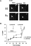
CaMKII activity is necessary for CEDE.A, KN-62 (5 μm) prevents CEDE. The 12 DIV culture, loaded overnight with FM1-43, was challenged with A23187 in the presence (top two panels) and after the washout (bottom two panels) of 5 μm KN-62, a CaMKII inhibitor. Scale bar, 20 μm. B, Plot of FM1-43 fluorescence observed in overnight-loaded mature neurons (>9 DIV) transiently challenged with A23187 (indicated by arrows) in the presence of 0.5 μm (dotted line;n = 3) or 5 μm KN-62 (solid line; n = 8; *p < 0.05) and after the subsequent washout of the drug.
Fig. 3.
Fig. 3.
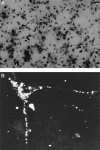
Infection of cultured neurons with the vaccinia virus. A, A large fraction (>90%) of cells in cultures exposed to a vaccinia virus (VV) producing recombinant β-galactosidase (βGALVV) expresses recombinant β-galactosidase, indicated by X-GAL staining. B, Expression of recombinant αCaMKII in a neuron infected with a VV producing both recombinant hemagglutinin epitope-tagged αCaMKII and β-galactosidase (fCaMKIIVV), indicated by punctate immunofluorescent staining with the use of a hemagglutinin epitope antibody.
Fig. 4.
Fig. 4.
Recombinant αCaMKII renders immature neurons competent to show CEDE. A, Neurons (7 DIV) loaded overnight with FM1-43 were uninfected (top) or were infected with βGALVV (middle) or CaMKIIVV (bottom). A23187 (1 μm for 1 min) evoked no significant exocytosis in uninfected and βGALVV-infected neurons but produced significant exocytosis in CaMKIIVV-infected neurons. Scale bar, 10 μm. B, Quantitative analysis of CEDE in control and infected developing neurons (age is indicated). The following number of cultures was analyzed for each experiment:5/6 DIV, control n = 12, βGALVVn = 7, CaMKIIVV n = 8;7/8 DIV, control n = 15, βGALVVn = 8, CaMKIIVV n = 9; 9 DIV, control n = 8, βGALVVn = 5, CaMKIIVV n = 5. *p < 0.05; **p < 0.01. At no age was CEDE significantly different in βGALVV and uninfected neurons.
Fig. 5.
Fig. 5.
Recombinant αCaMKII can be sufficient to produce dendritic exocytosis. A, Constitutively active αCaMKII (tCaMKII) produces dendritic exocytosis in the absence of a calcium stimulus and is blocked by nocodazole. Fluorescence images were obtained at time 0 (after 1 hr of wash, left panels) and 1 hr later (right panels) of cultures infected with βGALVV (top), tCaMKIIVV (middle), or tCaMKIIVV with nocodazole treatment (bottom). Scale bar, 10 μm. B, Fractional loss of fluorescence (indicating dendritic exocytosis) observed over 1 hr for the conditions indicated.Sample size in parentheses refers to the number of cultures examined. **p < 0.01.
Fig. 6.
Fig. 6.
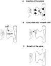
CEDE as a possible mechanism of synaptic plasticity. Ca2+-induced activation of αCaMKII leads to the regulated fusion of perisynaptic SER/TGN-derived organelles with the dendritic plasma membrane. This fusion could (A) expose new receptors at the surface of the postsynaptic cell, (B) release factors that might affect presynaptic function, or (C) provide more membrane for the growth of synapses.

Similar articles

Cited by

References

    1. Banker GA, Goslin K. Culturing nerve cells. MIT; Cambridge, MA: 1990.
    1. Barria A, Muller D, Derkach V, Griffith LC, Soderling TR. Regulatory phosphorylation of AMPA-type glutamate receptors by CaM-KII during long-term potentiation. Science. 1997;276:2042–2045. - PubMed
    1. Baude A, Nusser Z, Molnar E, McIlhinney RA, Somogyi P. High-resolution immunogold localization of AMPA-type glutamate receptor subunits at synaptic and non-synaptic sites in rat hippocampus. Neuroscience. 1995;69:1031–1055. - PubMed
    1. Bennett MK, Erondu NE, Kennedy MB. Purification and characterization of a calmodulin-dependent protein kinase that is highly concentrated in brain. J Biol Chem. 1983;258:12735–12744. - PubMed
    1. Braun AP, Schulman H. The multifunctional calcium/calmodulin-dependent protein kinase: from form to function. Annu Rev Physiol. 1995;57:417–445. - PubMed

Publication types

MeSH terms

Substances

LinkOut - more resources