. 1998 Sep 1;18(17):6871-81.
doi: 10.1523/JNEUROSCI.18-17-06871.1998.

Nicotinic receptor-induced apoptotic cell death of hippocampal progenitor cells

Affiliations

Nicotinic receptor-induced apoptotic cell death of hippocampal progenitor cells

F Berger et al. J Neurosci. .

Abstract

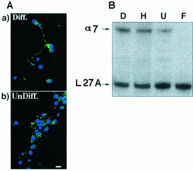
Nicotine has many effects on CNS functions, presumably through its action on neuronal nicotinic acetylcholine receptors (AChRs). One subclass of AChRs that binds the snake venom toxin alpha-bungarotoxin (alpha-Bgt-AChRs) has been shown to modulate neurotransmission in the brain. We now show that alpha-Bgt-AChR activation by low doses of nicotine results in apoptotic cell death of both primary and immortalized hippocampal progenitor cells. In HC2S2-immortalized hippocampal progenitors, nicotine is cytotoxic to undifferentiated cells, whereas it spares the same cells once differentiation has been induced. The activation of alpha-Bgt-AChRs by nicotine results in the induction of the tumor suppressor protein p53 and the cdk inhibitor p21. The cytotoxic effect of nicotine is dependent on extracellular calcium levels and is probably attributable to the poor ability of undifferentiated progenitors to buffer calcium loads. The major calcium buffer in these cells, calbindin D28K, is present only after differentiation has been induced. Furthermore transfection of undifferentiated cells with calbindin results in dramatic protection against the cytotoxic effects of nicotine. These results show that nicotine abuse could have significant effects on the survival of progenitor populations in the developing and adult brain and also suggest an endogenous role for alpha-Bgt-AChRs in neuronal development and differentiation.


Figures

Fig. 1.
Fig. 1.
Nicotine-induced cell death in primary hippocampal progenitors. Primary hippocampal progenitor cells were treated overnight with either control solution (Control) or 0.5 μm nicotine (Nicotine). Cell death was visualized using the Apoptag Kit (Oncor). Peroxidase-positive nuclei in cells that still retained their morphology were counted. Treatment with nicotine resulted in an increase in the number of apoptotic cells. Cell counts revealed a 30-fold increase in the number of Apoptag-positive cells after nicotine treatment (p < 0.005; Mann–Whitney Utest). This effect of nicotine was blocked by preincubation with 100 nm α-Bgt for 30 min, followed by its continued presence during nicotine treatment (Nic.+ αBgt). Five hundred cells per field, three fields per dish were counted. The values expressed in the y-axis are Apoptag-positive cells as a percentage of total cells counted from three separate experiments (mean ± SEM).
Fig. 2.
Fig. 2.
Effect of nicotine on the survival of HC2S2 progenitor cells. HC2S2 cells plated in 24 mm culture wells were treated with nicotine for a 36 hr period after which cell survival was assessed by trypan blue exclusion and counting. Values represent the mean ± SEM from two to four experiments, each done in triplicate.A, Bright-field images of HC2S2 cells that were untreated (a, Con), treated with 0.5 μmnicotine in the presence of 100 nm α-Bgt (b, Nic + Bgt), or treated with 0.5 μmnicotine alone (c, Nic). Nicotine caused a dramatic reduction in the number of trypan blue excluding cells; that reduction was completely reversed in the presence of α-Bgt. Scale bar, 50 μm.B, Quantitation of the cytotoxic effects of nicotine by cell counts. Nicotine-treated wells showed a 50% decrease in live cell numbers that was reversible by pretreating the cells with 100 nm α-Bgt followed by the continued presence of the toxin. The toxin by itself did not significantly affect cell numbers. Cell numbers in untreated control = 411 ± 59 × 103 cells/24 mm well (mean ± SEM from 4 experiments). Values are expressed as percent live cells compared with untreated controls (% Con). C, Dose–response for the effect of nicotine on the survival of undifferentiated HC2S2 cells. Nicotine significantly decreased the survival of undifferentiated progenitors at all the concentrations tested between 5 nm and 5 μm. The lesser efficacy of the 5 μm concentration could be attributable to a more rapid desensitization of the receptors. Values are mean ± SEM from two experiments done in triplicate.
Fig. 3.
Fig. 3.
Nicotine is not cytotoxic to differentiated HC2S2 cells. Differentiated HC2S2 cells were treated with 0.5 mmnicotine as described in Figure 2. Values represent the mean ± SEM from two experiments, each done in triplicate. A, Both control (Con) and nicotine-treated (Nic) cells showed robust neurons with well-defined processes. Scale bar, 50 μm. B, Cell counts from two independent determinations done in triplicate revealed no significant difference in cell numbers between the two conditions.
Fig. 4.
Fig. 4.
Presence of α-Bgt-AChRs on HC2S2 cells. α-Bgt-AChRs on HC2S2 cells were detected by surface radiolabeled toxin binding (see Results), fluorescence labeling, and RT-PCR experiments. A, Fluorescence labeling of HC2S2 cells. Cells were labeled with biotinylated α-Bgt followed by Cy3-conjugated extravidin. Background label was assessed by labeling in the presence of 1 μm α-Bgt. Both the differentiated (Diff) and the undifferentiated (Undiff) cells showed detectable levels of toxin binding on their surface (position of cells is represented by the DAPI staining in blue). In the differentiated cells, punctate labeling was also observed on the processes. Scale bar, 25 μm.B, RT-PCR showing the presence of α7 message in HC2S2 cells. Using primers flanking a 460 bp region of the putative cytoplasmic domain of the rat α7 gene, RT-PCR was performed on RNA isolated from differentiated HC2S2 cells (lane D), adult rat hippocampus (lane H), undifferentiated HC2S2 cells (lane U), and rat fibroblasts (lane F). The level of α7 message was normalized to that of the control ribosomal protein L 27A message in the same PCR mix. The α7 message is expressed in differentiated HC2S2 cells in levels comparable to those in the adult hippocampus. Lesser, although significant, expression was seen in the undifferentiated cells. Fibroblasts do not show any detectable expression of the α7 message.
Fig. 5.
Fig. 5.
Nicotine induces apoptosis in undifferentiated HC2S2 cells. The effect of treatment with 0.5 μm nicotine for 12 hr on the survival of undifferentiated HC2S2 cells was examined by the following methods. A, DNA laddering. DNA was extracted from cells 12 hr after no treatment (Con) or after treatment with 0.5 μmnicotine (Nic 0.5), 50 μmnicotine (Nic 50), or 0.5 μm nicotine in the presence of 100 nm α-Bgt (Nic +Bgt). Nicotine-induced DNA fragmentation characteristic of apoptosis was seen in undifferentiated HC2S2 cells and was prevented by pretreatment with α-Bgt. The arrow shows the position of the 247 bp DNA marker. B, Fluorescence and immunocytochemistry. Nicotine-induced cell death in undifferentiated HC2S2 cells is apoptotic as seen by nuclear fragmentation (arrow) visualized by staining undifferentiated cells with DAPI 12 hr after exposure to 0.5 μm nicotine (bottom panel). Immunofluorescence on the same field of cells indicates the induction of p53 (red) and p21 (green) or p53 and p21 (yellow) expression (top panel). In the presence of bungarotoxin, neither nuclear fragmentation nor the induction of p53/p21 is seen (insets). Scale bar, 25 μm. C, Western blotting. Western blots of untreated cells (lane 1,C), cells treated with 0.5 μm nicotine in the presence of 100 nm α-Bgt (lane 2,N + Bgt) or with nicotine alone at 0.5 μm (lane 3, N) revealed the induction of both p53 and p21 proteins by nicotine. No induction was observed in control cells or in cells treated with nicotine in the presence of α-Bgt. In differentiated HC2S2 cells (lanes 4, 5) no p53 signal was seen in either control (lane 4, C) or in cells treated with 0.5 μm nicotine (lane 5,N). Differentiated cells showed endogenous levels of p21 under both conditions and in a manner independent of the expression of p53. All lanes were probed with an actin antibody to estimate the approximate amounts loaded.
Fig. 6.
Fig. 6.
Nicotine-induced apoptosis is calcium-dependent. The effect of 0.5 μm nicotine on the survival of undifferentiated HC2S2 cells was examined at two different calcium concentrations (1.2 mm vs 100 μm). Lowering external calcium 10-fold to 100 μm reversed the ability of nicotine to mediate cell death in these cells. Cells were tested for nicotine-induced cytotoxicity in the presence or absence of 10 μm KN-62 (Calbiochem, La Jolla, CA), an inhibitor of CAM kinase II. In the presence of KN-62, the ability of nicotine to kill undifferentiated HC2S2 cells was reduced. The inhibitor by itself does not significantly affect cell survival. Results show that the effect of nicotine is calcium-dependent.
Fig. 7.
Fig. 7.
The apoptotic effects of nicotine are dependent on the expression of calbindin D28K. A, Expression of calbindin-D28K. Undifferentiated and differentiated HC2S2 cells were incubated overnight with Abs against calbindin D28K followed by an FITC-conjugated secondary. Little to no expression of calbindin was seen in undifferentiated HC2S2 cells, whereas robust signals were obtained from differentiated cells. These results suggest that calbindin is expressed only after the induction of differentiation in these cells. Scale bar, 25 μm. B, Rescue of undifferentiated HC2S2 cells by transient transfection of calbindin. Undifferentiated HC2S2 cells were transiently transfected with calbindin D28K. Cells were then treated with 0.5 μmnicotine and stained for nuclear fragmentation with DAPI (top panel) and with an anti-calbindin mAb (bottom panel). Calbindin-positive cells (top andbottom panels, long arrows) showed very little nuclear fragmentation, whereas cells that do not express the gene had large numbers of fragmented nuclei (top panel,short arrows). Scale bar, 25 μm. C, Quantitation of calbindin-mediated rescue. The numbers of fragmented nuclei were counted in undifferentiated cells treated with 0.5 μm nicotine that were calbindin-positive (D28 +ve) or did not express the gene (D28ve). Cells that did not express calbindin showed a 38-fold greater number of fragmented nuclei than calbindin-positive cells (p < 0.003, Mann–Whitney U test). Results are mean ± SEM from three independent determinations.

References

    1. Alkondon M, Rocha ES, Maelicke A, Albuquerque EX. Diversity of nicotinic acetylcholine receptors in rat brain. V: Alpha-bungarotoxin-sensitive nicotinic receptors in olfactory bulb neurons and presynaptic modulation of glutamate release. J Pharmacol Exp Ther. 1996;278:1460–1471. - PubMed
    1. Arends MJ, Harmon BV, Kerr JFR. Apoptosis: the role of the endonuclease. In: Potten CS, editor. Perspectives on mammalian cell death. Oxford UP; New York: 1990. pp. 229–258.
    1. Arneric SP, Sullivan JP, Decker MW, Brioni JD, Bannon AW, Briggs CA, Donnelly-Roberts D, Radek RJ, Marsh KC, Kyncl J, Williams M, Buccafusco JJ. Alzheimer’s disease and associated disorders, Vol 9, pp 50–61. Lippincott–Raven; Philadelphia: 1995. Potential treatment of Alzheimer’s disease using cholinergic channel activators (ChCAs) with cognitive enhancement, anxiolyric-like and cytoprotective properties. - PubMed
    1. Barrantes GE, Rogers AT, Lindstrom J, Wonnacott S. α-Bungarotoxin binding sites in rat hippocampal and cortical cultures: colocalization with α7 subunits and up-regulation by chronic nicotine treatment. Brain Res. 1995;672:228–236. - PubMed
    1. Beck KD, Hefti F, Widmer HR. Deafferentation removes calretinin immunopositive terminals, but does not induce degeneration of calbindin D-28k and parvalbumin expressing neurons in the hippocampus of adult rats. J Neurosci Res. 1994;39:298–304. - PubMed

Publication types