. 1998 Sep 1;18(17):6892-904.
doi: 10.1523/JNEUROSCI.18-17-06892.1998.

N-cadherin redistribution during synaptogenesis in hippocampal neurons

Affiliations

N-cadherin redistribution during synaptogenesis in hippocampal neurons

D L Benson et al. J Neurosci. .

Abstract

Cadherins are homophilic adhesion molecules that, together with their intracellular binding partners the catenins, mediate adhesion and signaling at a variety of intercellular junctions. This study shows that neural (N)-cadherin and beta-catenin, an intracellular binding partner for the classic cadherins, are present in axons and dendrites before synapse formation and then cluster at developing synapses between hippocampal neurons. N-cadherin is expressed initially at all synaptic sites but rapidly becomes restricted to a subpopulation of excitatory synaptic sites. Sites of GABAergic, inhibitory synapses in mature cultures therefore lack N-cadherin but are associated with clusters of beta-catenin, implying that they contain a different classic cadherin. These findings indicate that N-cadherin adhesion may stabilize early synapses that can then be remodeled to express a different cadherin and that cadherins systematically differentiate between functionally (excitatory and inhibitory) and spatially distinct synaptic sites on single neurons. These results suggest that differential cadherin expression may orchestrate the point-to-point specificity displayed by developing synapses.


Figures

Fig. 1.
Fig. 1.
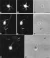
Localization and specificity of N-cadherin and β-catenin antibodies. A, Western blots of homogenates from 17-d-old cultured hippocampal neurons incubated with monoclonal antibody against N-cadherin (N-cad; left) or β-catenin (β-cat; right) are shown. Size standards are indicated on the left.B, C, Extracellular (B) and intracellular (C) domain-specific antibodies against N-cadherin colocalize in double-immunolabeled preparations of cultured hippocampal neurons.D, F, Preadsorbed polyclonal antibody yields no specific immunolabeling (F) compared with control (D). E,G, Corresponding phase-contrast photomicrographs are shown. Scale bars: B, C, 4 μm;D–G, 35 μm.
Fig. 2.
Fig. 2.
N-cadherin is present in axons and dendrites before synaptogenesis. Fluorescence photomicrographs showing N-cadherin (A, D, G) and synaptophysin (B, E) immunolabeling alongside corresponding phase-contrast photomicrographs (C, F, H) during early stages of neuron development are shown. Before axonal outgrowth, N-cadherin– (A) and synaptophysin- (B) labeled puncta are distributed throughout the cell soma and minor processes (C). After polarization, N-cadherin labeling (D) is detected in both the axon (arrow) and dendrites, whereas in the same neuron, synaptophysin labeling is polarized to the axon (E; arrow). Both labels are detected in soma (D, E). In G andH, N-cadherin is concentrated at the lamellipodial edge of a particularly large growth cone (arrow). Scale bars:A–F, 18 μm; G, H, 11 μm.
Fig. 3.
Fig. 3.
Synaptic localization of N-cadherin. Confocal images show N-cadherin (green) and synaptophysin (red) immunolabeling separately and overlaid in which regions of colocalization that appear yellow.A–C, In 5-d-old neurons, single optical sections show all N-cadherin–labeled puncta (A; green) contain synaptophysin label (B; red). When images are overlaid (C), the close association of N-cadherin and synaptophysin is readily apparent with each complex having a central area of colocalization (yellow). The synaptic complex (indicated by thearrowhead) is presented at higher magnification in each inset. D–F, By 14 d, most N-cadherin puncta (D; green) remain associated with synaptophysin boutons (E;red), but some synaptophysin boutons (arrow) lack N-cadherin (F). The larger size of green N-cadherin puncta and their relationship with red synaptophysin-labeled boutons can be seen in the higher magnification inset of the synaptic complex (indicated by the arrowhead) inD–F as well as in G andH. GH, N-cadherin appears to partially enclose synaptophysin labeling.I–J, Figures illustrate the differences between synaptophysin boutons that do not contain N-cadherin (arrows) but are close to those that are colocalized with N-cadherin (asterisks). Scale bars:A–C, 4 μm (insets, 1 μm);D–F, 11 μm (insets, 1 μm);G, 1 μm; H, 1.3 μm; I, 0.7 μm; J, 0.8 μm.
Fig. 4.
Fig. 4.
N-cadherin is lost from inhibitory synapses. Confocal images show low and high magnification images of N-cadherin (green) and GAD (purple) immunolabeling separately and overlaid where regions of colocalization appear white.A–D, At 7 d in culture, low (A) and high (B–D) magnification images show all GAD-labeled boutons associated with N-cadherin puncta. Synaptic complex indicated by anarrowhead is shown at higher magnification in eachinset (B–D) where a large area of colocalization appears white in overlay.E–H, In contrast, both low (E) and high (F–H) magnification images indicate that by 17 d in culture no GAD-labeled synapses contain N-cadherin. Scale bars: A, 25 μm;B–D, 5 μm (insets, 1 μm);E, 12 μm; F–H, 7 μm.
Fig. 5.
Fig. 5.
Rate of N-cadherin loss from GABAergic synaptic sites. The bar graph illustrates the percentage of GAD-labeled boutons containing N-cadherin label at different stages of development. The percentage drops sharply between 7 and 9 d and then continues to drop more slowly to nearly zero by 17 d in culture.
Fig. 6.
Fig. 6.
N-cadherin is associated with excitatory postsynaptic densities. Confocal images (A–C) and fluorescence photomicrographs (D, E) of cultured hippocampal neurons show colocalization of N-cadherin (D; green) with the excitatory postsynaptic markers PSD-95 (B, C;red) and GluR1 (E; red).A–C, In 29-d-old neurons, in single optical sections, most N-cadherin puncta cluster at PSD-95–containing postsynaptic densities, some of which are on dendritic spines. Those indicated byupper and lower arrowheads inA–C are shown at higher magnification in theupper and lower insets, respectively. D, E, N-cadherin puncta in 17-d-old neurons also associate with GluR1 clusters.Boxed regions are shown at higher magnification incolor (red, green, andoverlay) on the right where thearrowhead illustrates a double-labeled dendritic spine. Scale bars: A–C, 7.5 μm (insets, 1.7 μm); D, E, 18 μm, higher magnification, 6 μm.
Fig. 7.
Fig. 7.
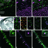
GABAergic synapses do not contain N-cadherin, but they do contain β-catenin. A–I, Confocal images and a bright-field photomicrograph of sections from adult brain double-labeled for N-cadherin (green) and GAD (purple) or synaptophysin (red) or stained for Nissl (D). Sections through the CA3 region of monkey hippocampus (A–C) show N-cadherin concentrated in mossy fiber terminals in stratum lucidum, whereas GABAergic terminals are concentrated in the pyramidal cell layer. Letters on Nissl-stained mouse brain sectionD correspond to regions from semiadjacent sections shown at high magnification in E–I. In single optical sections through layers II and III of cerebral cortex, GABAergic synapses (purple) do not contain N-cadherin (green) (E), but most N-cadherin puncta (green) are synaptophysin immunoreactive (red) and appear yellow(F). Differential distribution of GAD and N-cadherin is also observed in single optical sections through the habenula (G), hypothalamus (H), and caudate-putamen (I). J–L, Confocal images taken from 14-d-old cultured rat hippocampal neurons show β-catenin (green) and GAD (purple) immunolabeling separately (J, K) and overlaid (L) where regions of colocalization appear white. Each purple GAD-labeled bouton associates with green β-catenin–labeled clusters (e.g., arrows). The bouton indicated by thetop arrow is shown at high magnification in theinset, where the two labels can be seen to colocalize over a central white area. Scale bars:A–C, 17 μm; D, 1 mm; E,F, 7 μm; G–I, 7.5 μm;J–L, 5.6 μm (insets, 2.8 μm).
Fig. 8.
Fig. 8.
β-Catenin is found at synapses and in nuclei. A, B, Projection of confocal images through an entire cell somata (10 × 1 μm) indicates both surface β-catenin labeling and label within the nucleus (A), but projection of a subset of the same images (3 × 1 μm) emphasizes the nuclear distribution (B). The nucleolus is unlabeled, and the nuclear envelope appears to be labeled in a punctate manner. C,D, Fluorescence and phase photomicrographs show β-catenin–labeled puncta in both axons and young dendrites before synapse formation. Although not concentrated particularly in growth cones, puncta are observed occasionally in filopodia (insets). E, F, β-Catenin–labeled puncta (green) colocalize with synaptophysin-labeled (red) synapses as shown in phase and fluorescence photomicrographs of 13-d-old cultured neurons. Boxed regions indicated in E andF are shown at higher magnification on theright separately and overlaid.G–I, In confocal images of 26-d-old neurons, N-cadherin–labeled puncta (H; red) represent a subpopulation of β-catenin–labeled puncta (G; green). Most of the large β-catenin puncta correspond to those labeled for N-cadherin (G–I). Scale bars: A,B, 6 μm; C, D, 27 μm (insets, 6 μm); E, F, 20 μm (boxes, 10 μm); G–I, 2 μm.

References

    1. Amagai M, Karpati S, Prussick R, Klaus-Kovtun V, Stanley JR. Autoantibodies against the amino-terminal cadherin-like binding domain of pemphigus vulgaris antigen are pathogenic. J Clin Invest. 1992;90:919–926. - PMC - PubMed
    1. Apperson ML, Moon IS, Kennedy MB. Characterization of densin-180, a new brain-specific synaptic protein of the O-sialoglycoprotein family. J Neurosci. 1996;16:6839–6852. - PMC - PubMed
    1. Arndt K, Redies C. Restricted expression of R-cadherin by brain nuclei and neural circuits of the developing chicken brain. J Comp Neurol. 1996;373:373–399. - PubMed
    1. Arndt K, Nakagawa S, Takeichi M, Redies C. Cadherin-defined segments and parasagittal cell ribbons in the developing chicken cerebellum. Mol Cell Neurosci. 1998;10:211–228. - PubMed
    1. Bahr BA, Staubli U, Xiao P, Chun D, Ji Z-X, Esteban ET, Lynch G. Arg-Gly-Asp-Ser-selective adhesion and the stabilization of long-term potentiation: pharmacological studies and the characterization of a candidate matrix receptor. J Neurosci. 1997;17:1320–1329. - PMC - PubMed

Publication types