. 1998 Sep 1;18(17):6939-51.
doi: 10.1523/JNEUROSCI.18-17-06939.1998.

Functional magnetic resonance imaging of early visual pathways in dyslexia

Affiliations

Functional magnetic resonance imaging of early visual pathways in dyslexia

J B Demb et al. J Neurosci. .

Abstract

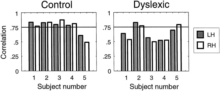
We measured brain activity, perceptual thresholds, and reading performance in a group of dyslexic and normal readers to test the hypothesis that dyslexia is associated with an abnormality in the magnocellular (M) pathway of the early visual system. Functional magnetic resonance imaging (fMRI) was used to measure brain activity in conditions designed to preferentially stimulate the M pathway. Speed discrimination thresholds, which measure the minimal increase in stimulus speed that is just noticeable, were acquired in a paradigm modeled after a previous study of M pathway-lesioned monkeys. Dyslexics showed reduced brain activity compared with controls both in primary visual cortex (V1) and in several extrastriate areas, including area MT and adjacent motion-sensitive areas (MT+) that are believed to receive a predominant M pathway input. There was a strong three-way correlation between brain activity, speed discrimination thresholds, and reading speed. Subjects with higher V1 and MT+ responses had lower perceptual thresholds (better performance) and were faster readers. These results support the hypothesis for an M pathway abnormality in dyslexia and imply strong relationships between the integrity of the M pathway, visual motion perception, and reading ability.


Figures

Fig. 1.
Fig. 1.
Psychometric function (percentage correct versus test speed) in a two-interval forced-choice speed discrimination experiment. Dashed line indicates the threshold speed increment that yielded 79% correct performance. Speeds are expressed as Weber fractions, i.e., as percentage increases over the baseline speed. Symbol size is proportional to the number of trials at a given test speed.
Fig. 2.
Fig. 2.
fMRI response variability. A, Average time series (averaged within MT+), from one subject for a 50% contrast moving grating stimulus, superimposed with the best fitting sinusoid (dashed line). B, Amplitude of Fourier transform of A. The signal frequency (6 cycles/scan) and its harmonics are represented by triangles. Filled circles correspond to nonharmonic frequencies. Solid curve is an exponential function fit that was used to estimate the noise amplitude at the signal frequency (dashed line).
Fig. 3.
Fig. 3.
fMRI response versus contrast. MT+ responses as a function of stimulus contrast in a dyslexic subject. Thecontinuous curve is a fitted power function, and the error bars represent estimates of the noise in the fMRI responses (see Materials and Methods). Dashed line indicates the fitted response at 20% contrast.
Fig. 4.
Fig. 4.
Top. Visual area locations. A, Parasagittal anatomical image from one subject indicating the slice selection (perpendicular to the calcarine sulcus) for the retinotopy measurements and the control conditions. B–D, Visual areas V1, V2, V3, V3A, and V4v depicted on in-plane anatomies from a similar location (near the middle of the 8 in-planes) in three control subjects. E–G, Visual areas depicted on in-plane anatomies from a similar location in three dyslexic subjects. Figure 5. Bottom. Location of area MT+. A, Parasagittal anatomical image from one subject indicating the slice selection (parallel to the calcarine sulcus) for the moving dots and the test conditions. The blue lines were slices that contained the MT+ region of interest (ROI) in this subject. B–D, Brain activity in one slice containing the MT+ ROI from three control subjects. Reddish voxels show regions with greater response to moving versus stationary dot patterns. Images were chosen to optimally show MT+ in the right hemisphere (arrows), although activity from left hemisphere MT+ and V1 are also present in some cases. The image in B is from the same brain as the sagittal image in A and corresponds to the most inferior of the three blue slices. The MT+ ROI was defined by outlining (dotted line in B) the strongest area of activity that was approximately lateral to the junction between the calcarine sulcus and the parieto-occipital sulcus, and beyond the retinotopically organized visual areas (see Materials and Methods). E–G, Slices with MT+ ROIs in three dyslexic subjects.
Fig. 6.
Fig. 6.
Differences in brain activity between dyslexic and control groups. Group average fMRI responses in MT+ and V1 to test stimuli (low mean luminance, moving gratings) as a function of stimulus contrast. Group differences were significant (p< 0.02) in both V1 and MT+ in these test conditions designed to preferentially stimulate the M pathway. Error bars represent ±1 SEM.Continuous curves are fitted power functions.
Fig. 7.
Fig. 7.
Bootstrapping statistical analysis. A, Bivariate distributions of bootstrapped power function parameters. The amplitude and exponent parameters for the 1000 bootstrapped contrast response functions are plotted for the dyslexic (x) and control (o) groups. Outliers from the dyslexic distribution (extreme exponent values) are omitted from the plot. B, The two bivariate distributions were projected onto an axis orthogonal to the best linear discriminator (dashed line) between the bivariate distributions. The resulting univariate distributions show a count of the points for dyslexic (gray) and control (white) groups. C, The difference distribution was created by randomly pairing the points from the projected univariate distributions twice and subtracting the dyslexic group values from the control group values. A final p value was derived by counting the number of values below zero (dashed line), after correcting for the sample size (Demb, 1997).
Fig. 8.
Fig. 8.
Similar brain activity in dyslexic and control groups. Group average fMRI responses in V1 to control stimuli (high mean luminance, contrast-reversing gratings) as a function of stimulus contrast. Group contrast responses were well matched (p > 0.10) in these control conditions designed to stimulate other pathways in addition to the M pathway. Error bars represent ±1 SEM. Continuous curves are fitted power functions. (MT+ activity was not recorded in this condition.)
Fig. 9.
Fig. 9.
Brain activity in four extrastriate areas.Top row, Group average fMRI responses in V2, V3, V3A, and V4v to test stimuli (low mean luminance, moving gratings) as a function of stimulus contrast. Control group responses were significantly greater than dyslexic group responses in all areas (p < 0.05). Bottom row, Group average fMRI responses to control stimuli (high mean luminance, contrast-reversing gratings) as a function of stimulus contrast. Dyslexic group responses were well matched or slightly higher in all areas. Error bars represent ±1 SEM. Continuous curves are fitted power functions.
Fig. 10.
Fig. 10.
Responses were measured to a moving dot stimulus that alternated with a gray field; stimulus parameters were similar to a previous fMRI study of dyslexia (Eden et al., 1996). After their analysis, the maximum correlation of all voxels in the MT+ ROI of each hemisphere is plotted for each subject. A line is drawn atr = 0.75 to demonstrate that under a given correlation threshold, the groups can be somewhat separated (8 of 10 control but only 3 of 10 dyslexic hemispheres above threshold), similar to the results reported by Eden et al. (1996).
Fig. 11.
Fig. 11.
Individual differences in both MT+ and V1 activity predict psychophysical speed discrimination thresholds. fMRI responses are the fitted values at 20% contrast. Psychophysical thresholds are averaged across three repeated measurements for each subject. Solid lines are regression lines through the data. Correlation between speed discrimination thresholds and MT+ activity was very strong (r = −0.79; p < 0.005). Correlation between speed thresholds and V1 activity was weaker, but still significant (r = −0.65;p < 0.025). Brain activity in V2, V3, V3A, and V4v was not correlated with speed discrimination performance.
Fig. 12.
Fig. 12.
Individual differences in brain activity are strongly correlated with individual differences in reading rate.Solid lines are regression lines through the data. The responses corresponding to contrasts that produced the highest correlations are shown (MT+, ct = 30%, r = 0.80; V1, ct = 85%, r = 0.68; V2, ct = 100%,r = 0.80; V3, ct = 100%, r = 0.77; V3A, ct = 53%, r = 0.60; V4v, ct = 100%, r = 0.80), although the correlations were significant across a wide range of contrasts in all areas (see Results). Reading rates are reported as percentile scores. The dyslexic subject with a high reading rate scored quite poorly on other reading measures, including the reading comprehension score of the Nelson-Denny reading test.

Similar articles

Cited by

References

    1. Albrecht DG. Visual cortex neurons in monkey and cat: effect of contrast on the spatial and temporal phase transfer functions. Vis Neurosci. 1995;12:1191–1210. - PubMed
    1. Beauchamp MS, Cox RW, DeYoe EA. Graded effects of spatial and featural attention on human area MT and associated motion processing areas. J Neurophysiol. 1997;78:516–520. - PubMed
    1. Beckers G, Homberg V. Cerebral visual motion blindness: transitory akinetopsia induced by transcranial magnetic stimulation of human area V5. Proc R Soc Lond B Biol Sci. 1992;249:173–178. - PubMed
    1. Beckers G, Zeki S. The consequences of inactivating areas V1 and V5 on visual motion perception. Brain. 1995;118:49–60. - PubMed
    1. Borsting E, Ridder WH, Dudeck K, Kelley C, Matsui L, Motoyama J. The presence of a magnocellular defect depends on the type of dyslexia. Vision Res. 1996;36:1047–1053. - PubMed

Publication types