. 1998 Sep;66(9):4208-14.
doi: 10.1128/IAI.66.9.4208-4214.1998.

Molecular cloning, expression, and immunogenicity of MTB12, a novel low-molecular-weight antigen secreted by Mycobacterium tuberculosis

Affiliations

Molecular cloning, expression, and immunogenicity of MTB12, a novel low-molecular-weight antigen secreted by Mycobacterium tuberculosis

J R Webb et al. Infect Immun. 1998 Sep.

Abstract

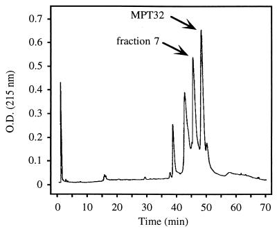
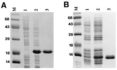
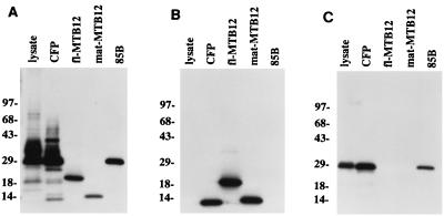
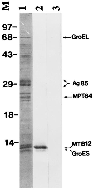
Proteins secreted into the culture medium by Mycobacterium tuberculosis are thought to play an important role in the development of protective immune responses. In this report, we describe the molecular cloning of a novel, low-molecular-weight antigen (MTB12) secreted by M. tuberculosis. Sequence analysis of the MTB12 gene indicates that the protein is initially synthesized as a 16.6-kDa precursor protein containing a 48-amino-acid hydrophobic leader sequence. The mature, fully processed form of MTB12 protein found in culture filtrates has a molecular mass of 12. 5 kDa. MTB12 protein constitutes a major component of the M. tuberculosis culture supernatant and appears to be at least as abundant as several other well-characterized culture filtrate proteins, including members of the 85B complex. MTB12 is encoded by a single-copy gene which is present in both virulent and avirulent strains of the M. tuberculosis complex, the BCG strain of M. bovis, and M. leprae. Recombinant MTB12 containing an N-terminal six-histidine tag was expressed in Escherichia coli and purified by affinity chromatography. Recombinant MTB12 protein elicited in vitro proliferative responses from the peripheral blood mononuclear cells of a number of purified protein derivative-positive (PPD+) human donors but not from PPD- donors.


Figures

FIG. 1
FIG. 1
Chromatogram of M. tuberculosis CFP fractionated by conventional HPLC (C18) followed by microbore HPLC (C18) and eluted with a 20 to 70% acetonitrile gradient. Peak fractions were analyzed by N-terminal amino acid sequencing. The peak labeled fraction 7 was found to contain a protein with novel N-terminal sequence. The peak labeled MPT32 was found to correspond to the 45/47-kDa secreted antigen (21).
FIG. 2
FIG. 2
(A) Nucleotide sequence and predicted amino acid sequence of the M. tuberculosis MTB12 gene. Those amino acids comprising the putative leader sequence (absent from the mature protein) are shown in italics. The amino acid residues determined during N-terminal sequence analysis of HPLC fraction 7 are shown in bold. The position of the β-galactosidase–MTB12 fusion in clone Ra-1 (isolated during a library screen using anti-CFP polyclonal antiserum) is indicated by an arrow at position 64. (B) Comparison of the amino acid sequence of the M. tuberculosis MTB12 protein and the predicted protein sequence of the M. leprae homolog (GenBank accession no. U00016_13). Identical amino acids are shown with a shaded background. The amino acid residues determined during N-terminal sequence analysis are indicated by a horizontal line above the MTB12 sequence.
FIG. 3
FIG. 3
Expression and purification of rMTB12 proteins. Recombinant proteins corresponding to full-length MTB12 protein (A) or mature MTB12 protein lacking the leader sequence (B), both containing amino-terminal six-histidine tags, were expressed by using the pET17b E. coli high-level expression system. Lysates of uninduced cultures (lane 1), IPTG-induced cultures (lane 2), and affinity-purified recombinant protein (lane 3) were fractionated by SDS-PAGE and stained with Coomassie blue. Positions of size markers (lane M) are indicated in kilodaltons.
FIG. 4
FIG. 4
Immunoblot analyses of MTB12. Western blots containing M. tuberculosis lysate (2.5 μg), CFP (2.5 μg), recombinant mature MTB12 (50 ng), full-length MTB12 (Fl-MTB12; 50 ng), and 85B (50 ng) were incubated with anti-CFP (A), anti-MTB12 (B), or anti-85B (C) polyclonal rabbit antisera. Immunoreactive proteins were detected by using [125I]protein A followed by autoradiography. Positions of size markers are indicated in kilodaltons.
FIG. 5
FIG. 5
Relative abundance of MTB12 in M. tuberculosis culture filtrates. M. tuberculosis CFP was separated by SDS-PAGE, transferred to a PVDF membrane by Western blotting and stained with Coomassie blue (lane 1). Bands corresponding to the most highly abundant proteins were excised and characterized by N-terminal amino acid sequencing. Identities of these proteins are indicated where known. A pair of duplicate strips were destained in 100% methanol, blocked with 1% bovine serum albumin and probed with a rabbit polyclonal anti-MTB12 antiserum (lane 2) or corresponding preimmune serum (lane 3). The middle band of the triplet migrating at approximately 12 kDa was shown by both amino acid sequencing and Western blot analysis to be MTB12. Positions of size markers (M) are indicated in kilodaltons.

References

    1. Aldrich C J, De Cloux A, Woods A S, Cotter R J, Soloski M J, Forman J. Identification of a Tap-dependent leader peptide recognized by alloreactive T cells specific for a class Ib antigen. Cell. 1994;79:649–658. - PubMed
    1. Andersen P. Effective vaccination of mice against Mycobacterium tuberculosis infection with a soluble mixture of secreted mycobacterial proteins. Infect Immun. 1994;62:2536–2544. - PMC - PubMed
    1. Andersen P, Askgaard D, Ljungqvist L, Bennedsen J, Heron I. Proteins released from Mycobacterium tuberculosis during growth. Infect Immun. 1991;59:1905–1910. - PMC - PubMed
    1. Boesen H, Jensen B N, Wilcke T, Andersen P. Human T-cell responses to secreted antigen fractions of Mycobacterium tuberculosis. Infect Immun. 1995;63:1491–1497. - PMC - PubMed
    1. Braud V, Jones E Y, McMichael A. The human major histocompatibility complex class Ib molecule HLA-E binds signal sequence-derived peptides with primary anchor residues at positions 2 and 9. Eur J Immunol. 1997;27:1164–1169. - PubMed

Publication types

MeSH terms

Associated data