. 1998 Sep;66(9):4274-82.
doi: 10.1128/IAI.66.9.4274-4282.1998.

Deletion of the central proline-rich repeat domain results in altered antigenicity and lack of surface expression of the Streptococcus mutans P1 adhesin molecule

Affiliations

Deletion of the central proline-rich repeat domain results in altered antigenicity and lack of surface expression of the Streptococcus mutans P1 adhesin molecule

L J Brady et al. Infect Immun. 1998 Sep.

Abstract

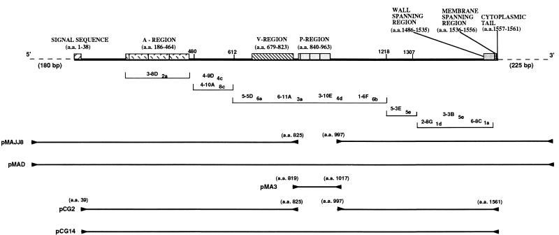
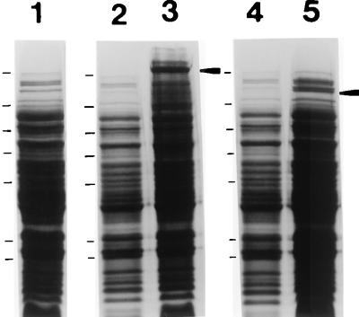
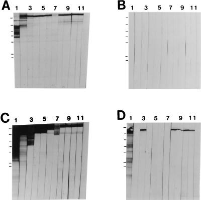
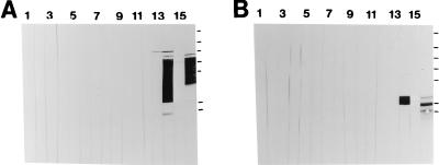
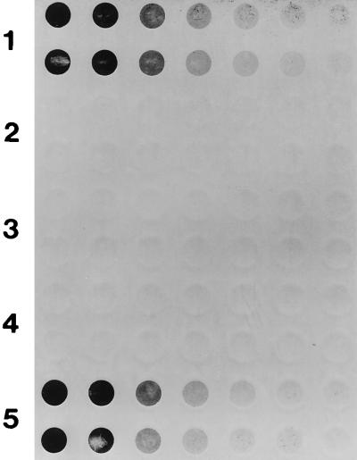
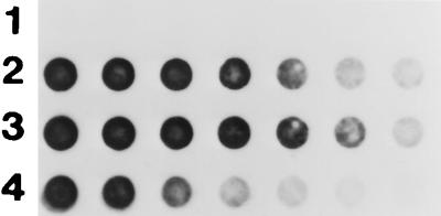
Members of the family of surface adhesins of oral streptococci, including P1 of Streptococcus mutans, contain two highly conserved repeat domains, one rich in alanine (A region) and the other rich in proline (P region). To assess the contribution of the P region to the biological properties of P1, an internal deletion in spaP was engineered. In addition, the P region was subcloned and expressed as a fusion partner with the maltose binding protein of Escherichia coli and liberated by digestion with factor Xa. Results of Western blot experiments in which recombinant polypeptides were probed with a panel of 11 monoclonal antibodies indicated that the P region is a necessary component of conformational epitopes within the central portion of P1. Antibodies reactive with the P region were detected in a polyclonal rabbit antiserum generated against whole S. mutans cells but not in two rabbit antisera generated against purified P1 (Mr approximately 185,000), suggesting that this domain is immunogenic on the surface of intact bacteria but not as part of a soluble full-length molecule. Finally, transformation of a spaP-negative mutant with a shuttle vector containing an internally deleted spaP lacking P-region DNA resulted in a complete absence of surface-localized P1 and substantially less P1 in sonicated cells compared to the case for the mutant complemented with the full-length gene. These results suggest that the P region is an integral component contributing to the conformation of the central region of P1 and indicate that its presence is necessary for surface expression of the molecule on S. mutans.


Figures

FIG. 1
FIG. 1
Schematic representation of the linear structure of P1. A linear map showing salient features of P1 is at the top. Approximate binding sites of the anti-P1 MAbs 3-8D2a, 4-10A8c, 4-9D4c, 5-5D6a, 6-11A3a, 3-10E4d, 1-6F6b, 5-3E5e, 2-8G1d, 3-3B5e, and 6-8C1a are shown below the linear map. The order in which these antibodies are listed within each indicated segment is arbitrary and does not reflect their exact binding locations. The locations of the forward and reverse PCR primers used to amplify spaP DNA are indicated by forward and reverse arrowheads, while the amplified DNA sequences contained in the indicated plasmids are represented by solid lines in the bottom half. Additional information regarding these plasmids is given in Table 1. a.a., amino acids.
FIG. 2
FIG. 2
SDS-polyacrylamide gel electrophoresis of E. coli containing plasmids with internally deleted and full-length spaP. Lane 1, cell lysate following IPTG induction of E. coli M15(pREP4) containing the His-tagged pQE30 vector only. Lanes 2 and 3, lysates of CG14 (full-length spaP) before and after IPTG induction, respectively. Lanes 4 and 5, lysates of CG2 (spaP with the P region deleted) before and after IPTG induction, respectively. The IPTG-induced P1 products are indicated by arrowheads to the right of lanes 3 and 5. The migrations of molecular weight standards are indicated to the left of lanes 1, 2, and 3 and correspond to Mrs of 180,000, 116,000, 84,000, 58,000, 48,500, 36,000, and 26,500.
FIG. 3
FIG. 3
Western blot analysis of recombinant full-length P1 and P1 lacking the P region. Lanes 1 through 11, reaction with anti-P1 MAbs 3-8D2a, 4-10A8c, 4-9D4c, 5-5D6a, 6-11A3a, 3-10E4d, 1-6F6b, 5-3E5e, 2-8G1d, 3-3B5e, and 6-8C1a, respectively (Fig. 1). Lane 12, reaction with the isotype-matched negative control MAb directed against A. actinomycetemcomitans. (A) Recombinant P1 purified from clone SMI/II (46). (B) Cell lysate of E. coli M15(pREP4) containing the His-tagged vector pQE30 only. (C) Cell lysate of CG14 (full-length spaP). (D) Cell lysate of CG2 (spaP with the P region deleted). The migrations of molecular weight standards are indicated to the left of lane 1 in each panel and correspond to Mrs of 180,000, 116,000, 84,000, 58,000, 48,500, 36,000, and 26,500.
FIG. 4
FIG. 4
Western blot analysis of the P region of P1. Lanes 1 through 11, reaction with MAbs 3-8D2a, 4-10A8c, 4-9D4c, 5-5D6a, 6-11A3a, 3-10E4d, 1-6F6b, 5-3E5e, 2-8G1d, 3-3B5e, and 6-8C1a, respectively (Fig. 1). Lane 12, reaction with the negative control anti-A. actinomycetemcomitans MAb 1-5F2a. Lane 13, reaction with mouse anti-β-galactosidase. Lane 14, reaction with rabbit anti-MBP. Lane 15, reaction with rabbit antiserum 209 made against purified P1 from S. mutans. Lane 16, reaction with rabbit antiserum 218 made against NG8 cells. (A) MBP–P-region fusion polypeptide recovered from recombinant E. coli periplasmic fraction by amylose resin chromatography. (B) MBP–P-region fusion polypeptide digested with factor Xa. The migrations of molecular weight standards are indicated to the right of lane 16 in each panel and correspond to Mrs of 180,000, 116,000, 84,000, 58,000, 48,500, 36,000, and 26,500.
FIG. 5
FIG. 5
Dot blot analysis of P1 expression by S. mutans NG8, spaP-negative mutant PC3370, and derivatives. Twofold serial dilutions of bacterial cells, beginning at 5 × 10⁶ CFU/well, were applied in duplicate to the membrane. Rabbit antiserum 209 made against purified P1 from S. mutans was used as the primary antibody. Rows 1 through 5, NG8, spaP-negative mutant PC3370, PC3370A (shuttle vector only), PC3370B (spaP with the P region deleted), and PC3370C (full-length spaP), respectively.
FIG. 6
FIG. 6
Western blot analysis of P1 expression by S. mutans NG8, spaP-negative mutant PC3370, and derivatives. Lanes 1 through 5, sonicated cell extracts prepared from NG8, PC3370C (full-length spaP), PC3370B (spaP with the P region deleted), PC3370A (shuttle vector only), and PC3370 with no plasmid, respectively. The filter was reacted with a cocktail of MAbs including 3-8D2a, 4-9D4c, 5-3E5e, 2-8G1d, 3-3B5e, and 6-8C1a (Fig. 1 and 3). The migrations of molecular weight standards are indicated to the left of lane 1 and correspond to Mrs of 180,000, 116,000, 84,000, 58,000, and 48,500. Arrowheads (from top to bottom) indicate full-length and internally deleted P1.
FIG. 7
FIG. 7
RNA dot blot analysis of spaP-specific mRNA levels in spaP-negative mutant PC3370 derivatives. Twofold serial dilutions of total cellular RNA, beginning with 5 mg, were probed with DNA encoding the A region of spaP. Rows 1 through 3, PC3370A (shuttle vector only), PC3370C (full-length spaP), and PC3370B (spaP with the P region deleted), respectively. Row 4, twofold serial dilutions of purified pMAD (Table 1), beginning with 100 ng.

References

    1. Ackermans S F, Klein J P, Ogier J A, Bazin H, Cromont F, Franck R M. Purification and characterization of a saliva interacting cell wall protein from Streptococcus mutans serotype f by using monoclonal antibody immunoaffinity chromatography. Biochem J. 1985;228:211–217. - PMC - PubMed
    1. Applied Biosystems, Inc. User bulletin 18. Piscataway, N.J: Applied Biosystems, Inc., Pharmacia Biotech; 1991.
    1. Ayakawa G Y, Boushell L W, Crowley P J, Erdos G W, McArthur W P, Bleiweis A S. Isolation and characterization of monoclonal antibodies specific for antigen P1, a major surface protein of mutans streptococci. Infect Immun. 1987;55:2759–2767. - PMC - PubMed
    1. Bleiweis A S, Karakawa W W, Krause R M. Improved technique for preparation of streptococcal cell walls. J Bacteriol. 1964;88:1198–1200. - PMC - PubMed
    1. Brady L J, Crowley P J, Ma J K-C, Kelly C, Lee S F, Lehner T, Bleiweis A S. Restriction fragment length polymorphisms and sequence variation within the spaP gene of Streptococcus mutans serotype c. Infect Immun. 1991;59:1803–1810. - PMC - PubMed

Publication types

MeSH terms

LinkOut - more resources